
Page 36 - AN-2-1
P. 36
Advanced Neurology The anterior cingulate cortex in social empathy
and periaqueductal gray matter predicts a longitudinal
increase in empathic concern in AD patients. These
results suggest that the acquisition of empathic concern
may be a very early feature of AD pathophysiology,
involving hyperconnectivity in a system that supports
emotion generation and perception [101] . Therefore, an early
identification of changes in empathic abilities may help in
the early detection of neuropsychiatric disorders in some
patients.
Patients with autism spectrum disorder (ASD) tend to
exhibit reduced empathic abilities and impaired emotional
empathy [102,103] . Imaging studies have demonstrated
structural defects in the core empathic network in autism,
such as reduced volumes of gray matter in bilateral superior
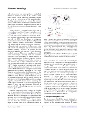
temporal gyri (STG) and disturbances in resting-state Figure 2. Neural circuits of ACC neurons projecting to the neural nuclei
functional connectivity between the ACC and prefrontal in pain and fear empathy. When empathy mice observe stimulated mice
lobes . However, another study has shown that patients receiving click and mechanical tingling stimuli through visual, olfactory,
[99]
with autism lack the ability to recognize, understand, and auditory sensory organs, there is activation of ACC neurons projecting
to downstream nuclei through the stimulation of ACC-BLA, ACC-MDL,
and describe their own emotions. On that account, while and ACC-MeA neural circuits as well as ACC internal microcircuits
ASD patients with alexithymia may experience emotional in fear empathic behaviors (blue arrows), while the ACC-NAc and the
empathy differently, it should not be described as a lack ACC-LC neural circuits mediate pain empathic behaviors (red arrows).
of ability to empathize with emotions [104] . Since there is no The neural circuit from the ACC to the LC has been proposed to mediate
standard, consistent definition of empathy, controversial pain empathy.
ACC: Anterior cingulate cortex; BLA: Basolateral nucleus of amygdala;
views have emerged. Although there are consistent Cg1/2: Cingulate cortex 1/2; LC: Locus coeruleus; MDL: Mediodorsal
assessment scales and evidence of reduced empathy in thalamic nucleus; MeA: Medial amygdala; NAc: Nucleus accumbens.
people with autism from the data on Empathy Quotient
(EQ), a 60-item self-report measure , the questions in social perception and behavioral understanding [112] .
[8]
such measures may be vague and imprecise as it is not Therefore, further investigations are required to determine
clear to whom or which group one should be comparing whether oxytocin contributes to prosocial behavior by
with [105] . Further grouping of individuals with autism, acting on ACC mirror neurons. In addition, it has been
and thus individualizing treatment, will be necessary. proposed that oxytocin release is potentially environment-
The use of different behavioral paradigms combined with dependent and might be involved in stress response, with
neurophysiological markers measured by fMRI-based a potential synchronous activation with cortisol; however,
blood oxygen level dependent (BOLD) response for the the two hormones are secreted independently, and
assessment of ASD patients might be a good way [106] . As stressor exposure is not the primary trigger for oxytocin
mentioned earlier, the MNS plays an important role in secretion [113,114] . Oxytocin may bind to postsynaptic
the perception of emotions and the imitative behaviors of oxytocin receptors in fast-spiking interneurons in the ACC,
individuals with ASD, which would further help to mediate significantly decreasing the action potential threshold and
empathic behaviors [107] . Future studies examining the resting membrane potential of interneurons, promoting the
effects of specific treatments on specific neurophysiological depolarization of interneurons, resulting in a decrease in
markers of MNS may reveal diagnostic subgroups of the E/I ratio, as well as enhancing the input from inhibitory
patients with ASD, with each subgroup requiring a specific to pyramidal neurons . The above studies suggest that the
[93]
treatment approach [106] . modulatory neural circuitry of oxytocinergic projections
Central or peripheral oxytocin injections are clinically to the ACC from other brain regions or subcortical nuclei
used to treat diminished empathic abilities in patients may be involved in the treatment of neuropsychiatric
with neuropsychiatric disorders [108-110] . Previous studies patients with impaired empathic abilities.
in rodent models of observational fear have shown that
intranasal oxytocin enhanced freezing behavior following 7. Perspective significance
observational fear transmission [54,111] . Recent studies The development of fMRI has greatly facilitated the
have also shown that intranasal oxytocin administration understanding of the neural basis behind empathy and
might act on the MSN in the sensorimotor circuit of depicted a vast neurofunctional network of various
Volume 2 Issue 1 (2023) 9 https://doi.org/10.36922/an.281