
Page 36 - AN-3-4
P. 36
Advanced Neurology Anticoagulants as neuroprotective therapeutics
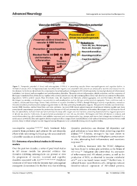
Figure 2. Therapeutic potential of direct oral anticoagulants (DOACs) in preventing vascular-driven neuropathogenesis and cognitive decline in
Alzheimer’s disease (AD). In hippocampal and neocortical brain regions, toxic amyloid-ß (Aß) proteins are produced by neurons and released into the
bloodstream. In the blood, Aβ activates the contact system by activating blood clotting factor XII, which leads to the increased production of inflammatory
bradykinin (not shown) and procoagulant and proinflammatory thrombin. Thrombin induces inflammation, platelet activation, and the conversion of
fibrinogen to insoluble fibrin. Fibrin forms, together with activated platelets and Aβ, Aβ-containing fibrin (fibrin-Aβ) clots that are resistant to fibrinolytic
degradation. Within cerebral vessels, these thrombotic clots, Aβ oligomers and filaments, platelet aggregates, and proinflammatory thrombin and
fibrin(ogen) accumulate, contributing to vasoconstriction and vasculopathies, such as Aβ-type cerebral amyloid angiopathy. These conditions result in
occlusive, cellular, and hemorrhagic lesions. Early inhibition of excessive thrombin by DOACs, through blocking its activity or production, normalizes
thrombin-mediated proinflammatory and procoagulant states in AD, thus preventing vasculopathic sequelae. This preserves vascular and blood–brain-
barrier (BBB) function, cerebral blood flow, and brain perfusion. An intact BBB ensures proper transport of substances between vascular and brain
parenchymal compartments, promotes the perivascular clearance of Aβ, and prevents the infiltration of thrombin, fibrin(ogen), and Aβ from the blood
into the parenchyma. Sufficient brain perfusion provides the tissue with oxygen and vital nutrients, preventing hypoxia/ischemia-triggered generation
of Aβ and tau proteins. Collectively, the parenchymal accumulation of toxic Aβ, tau, thrombin, fibrin(ogen), and fibrin-Aβ deposits is reduced, and
neuroinflammatory (e.g., glial activation and oxidative responses) and neurodegenerative (e.g., synapse and neuron loss) changes are minimized. As a
result, neural network function and cognitive abilities are preserved for a longer period. Early inhibition of Aβ-induced thrombin activity by DOACs could
prevent, delay, or at least mitigate vascular-triggered neuropathogenesis in AD, potentially slowing overall cognitive decline.
vasculopathic sequelae. 15,16,22-25,27 Early treatment helps thrombin production can decrease Aβ deposition and
preserve brain perfusion and optimize the anti-dementia glial activation in brain tissue while preserving cognitive
effects while minimizing the bleeding risks associated with abilities. 102,103 Likewise, enoxaparin has been shown to
vulnerable vasculature in elderly patients. reduce Aβ-induced activation of the plasma contact system
in vitro, attenuating both inflammatory and neurotoxic
5.1. Outcomes of preclinical studies in AD mouse responses. 102
models
In addition, treatment with the DOAC dabigatran
Over the past two decades, a series of preclinical studies has been found to reduce glial activation in the brains of
in AD mouse models has provided evidence that AD mice. Dabigatran also diminished the expression
122
thrombin-inhibiting anticoagulants can prevent or slow of inflammatory and AD-related proteins, as well as the
the progression of vascular, neuronal, and cognitive production of ROS, as observed in vascular endothelial
disorders associated with AD. 90,101-103,105,111,122,123 Peripheral cells and a tau-based mouse model. Furthermore, in
105
123
treatment of AD mice with the heparin-type anticoagulant cultured human neuroblastoma cells, dabigatran reduced
enoxaparin demonstrated for the first time that inhibiting thrombin-induced expression of AD-related proteins,
Volume 3 Issue 4 (2024) 18 doi: 10.36922/an.3799