
Page 112 - AN-4-1
P. 112
Advanced Neurology Dysarthria with CSF overdrainage syndrome
showing pressures over 200 mm of water; however, relief upper and lower extremity tremors, coldness in the left leg,
from this procedure is usually temporary. Treatment dizziness, tinnitus, blurry vision, and new-onset staccato
options include weight loss, corticosteroids, acetazolamide, speech (ataxic dysarthria) with stuttering followed by
and furosemide. 3 aphasia. Symptoms were triggered by standing and episodes
The most common treatment for IIH and idiopathic of whole-body shaking were relieved by lying down.
normal pressure hydrocephalus is a ventriculoperitoneal Physical examination revealed terminal intention tremors,
(VP) shunt. Complications of VP shunts include dysdiadochokinesia, dysmetria, difficulty with the heel-to-
infection, improper catheter placement, intraventricular shin test, left nystagmus, and mild leg spasticity, but cranial
hemorrhage, ventricular collapse, subdural hematoma, nerve function was normal with no other focal deficits.
4
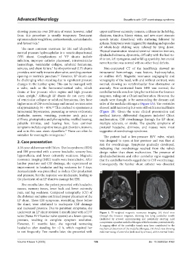
seizures, and shunt failure. Lumboperitoneal (LP) shunts Non-contrasted CT of the head/brain showed no
provide a minimally invasive alternative, avoiding cranium intracranial hemorrhage, mass lesions, hydrocephalus,
opening or ventricle puncture. However, LP shunts can or midline shift. Magnetic resonance angiography and
5,6
be challenging when standing due to significant pressure venography of the head, with and without contrast, were
changes in the lumbar spine. This can be managed with normal, showing no vertebrobasilar flow obstruction or
a valve, such as the horizontal-vertical valve, which anomaly. Non-contrasted brain MRI was normal; the
drains at low pressure when supine and high pressure cerebellar tonsils were low-lying but not below the foramen
when upright. Although LP shunts do not carry risks magnum, ruling out a Chiari malformation. However, the
7
of ventricular collapse or subdural hematoma, they have tonsils were thought to be surrounding the dorsum and
higher rates of CSF overdrainage and annual revision rates sides of the medulla oblongata (Figure 1A). The ventricles
of approximately 34 – 40%. This can lead to spontaneous showed mild narrowing but were still within normal limits
8,9
intracranial hypotension, presenting as severe orthostatic (Figure 1B). Given the acute clinical presentation and
headache, nausea, vomiting, posterior neck pain or medical history, differential diagnoses included Chiari
stiffness, photophobia and phonophobia, muffled hearing, malformation, CSF overdrainage through the LP shunt,
pulsatile tinnitus, and hearing loss. Less common multiple sclerosis, or stroke. The specific symptoms of
symptoms include cognitive issues, gait disorders, tremors, postural headache, dizziness, and nausea were most
and as in this case. ataxic dysarthria. These can often be suggestive of overdrainage syndrome.
10
mistaken for meningitis or migraine. 2 The patient had a low-pressure H/V valve, which
2. Case presentation was designed to open at low pressure and was thus at
risk for overdrainage. Symptoms gradually developed,
A 38-year-old woman with Ehlers–Danlos syndrome (EDS) indicating that overdrainage resulted from the valve’s
and IIH presented with a severe headache, sensory loss, design rather than shunt malfunction. The presence of
hyperreflexia, and lower extremity weakness. Magnetic dysdiadochokinesia and other cerebellar signs suggested
resonance imaging (MRI) results were inconclusive. After that the cerebellar tonsils sagged due to CSF overdrainage.
lumbar puncture and CSF drainage, she experienced an Consequently, the lumbar shunt catheter was dissected
improvement in headache and leg weakness for 3 days.
Acetazolamide was prescribed to reduce CSF production A B
and pressure, but the response was inadequate, leading to
the placement of an LP shunt to manage her IIH.
Five months later, the patient presented with headache,
nausea, memory issues, lower back and lower extremity
pain, and leg weakness. Computed tomography (CT) of
the abdomen and spine confirmed proper placement of the
LP shunt. These IIH symptoms, resembling those before
the shunt, were attributed to inadequate CSF drainage
and increased pressure. Due to persistent symptoms, she
underwent an LP shunt revision 1 month later with a H/V Figure 1. T1-weighted magnetic resonance imaging. (A) Axial view
valve (Natus H-V lumbar valve system) at a lower opening through the foramen magnum, showing low lying cerebellar tonsils
pressure, resulting in complete symptom resolution. (indicated by arrows) approximating and potentially exerting mild
However, 11 months later, she reported worsening compression upon the medulla oblongata of the brainstem posterolaterally.
A sagging effect of the cerebellar tonsils may have contributed to the
headaches after standing for >2 h, which required her mechanical distortion of the medulla oblongata. (B) Axial view showing
to rest frequently. Two months later, she presented with mild narrowing of ventricles (indicated by arrows), within normal limits
Volume 4 Issue 1 (2025) 106 doi: 10.36922/an.4162