
Page 34 - ARNM-2-3
P. 34
Advances in Radiotherapy
& Nuclear Medicine Molecular imaging of lung cancer
interventions such as surgery resection, chemotherapy, skeletal muscle, and other organs. CT scanning has been
and radiotherapy. 6,10,11 Therefore, the detection of extensively used as the benchmark reference for validating
subclinical lung cancer and metastases is essential for alternative field methods in human body compartment
timely treatment, reducing reoccurrence, and improving evaluation. By comparing the topographical images
35
patient stratification. obtained from CT scans to cross-sectional areas of
Surgical lung biopsies, the pathological gold standard tissues, data show high accuracy and reproducibility in
for early-stage lung cancer, are very challenging in patients distinguishing healthy tissue from tumor tissue in mouse
36
with small nodules. These procedures are invasive, models with induced tumors. Routine CT scans are
prone to high sampling errors, and often associated applied as a quantitative tool for detecting and monitoring
with complications. Computed tomography (CT), the lung cancer in both clinical settings and preclinical
12
radiological gold standard, is commonly utilized for animal studies. Major findings show the efficacy of CT
early lung cancer detection. 6,10,11 However, CT’s accuracy in detecting lung cancer at an early stage, especially for
in detecting malignant tumors is limited, as it provides adenocarcinoma. 37
minimal molecular-level information about tumor 2.1. Micro-computed tomography (micro-CT) and
microenvironment (TME) changes related to disease low-dose CT for non-invasive lung cancer imaging
invasion, progression, and regression. 13-15 There is an
urgent, unmet medical need for early and noninvasive High-resolution micro-CT has emerged as a powerful
detection of subclinical lung cancer and metastasis, as well tool for non-invasively visualizing and monitoring lung
as for monitoring their progression and regression. This cancer development in live mice. Low-dose computed
review summarizes current clinical and preclinical studies tomography (LDCT) has been effectively applied for lung
to highlight progress (Table 1) and challenges in molecular cancer screening in humans. CT is effective at both early
imaging for the early detection of lung cancer, along with and advanced stages of the disease, as validated against
their targeting mechanisms. traditional histological analysis. Notably, micro-CT can
distinguish lung tumors as small as 200 µm from healthy
2. CT tissue in a mouse model with induced tumors. This
remarkable capability is achieved with a low radiation dose
CT scans have long been applied to produce high- 38
resolution cross-sectional images, enabling the non- (0.4 Gy/15 min) and high spatial resolution (15 µm).
invasive separation, screening, and quantitative assessment CT primarily targets high-risk patients, placing a
of various tissue types, including bones, adipose tissue, significant burden on health-care systems. Although
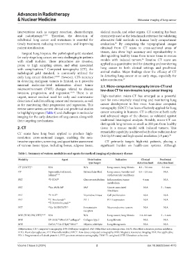
Table 1. Summary of various modalities and agents for medical imaging of pulmonary disease
Modality Agent Distribution Indication Clinical Preclinical
type/target detection limit detection limit
CT (LDCT) 16 N/A N/A Lung cancer; lung fibrosis 4.0 – 5.0 mm 0.2 mm
CT Iopromide; Iodixanol; Extracellular fluid Lung cancer; vascular and 0.5 – 2.0 mm N/A
Iohexol 17-19 cardiovascular conditions
PET 18 F-FDG 20 Glucose metabolism Inflammation; tumor 4 mm N/A
metabolism
PET 68 Ga-FAPI-04 21 FAP Cancer-associated N/A 3 – 5 mm
fibroblasts
PET 18 F-FLT 22 Thymidine kinase Cell proliferation N/A N/A
PET 89 Z-Nivolumab 23 PD-1 PD-1 expression N/A N/A
89 Z-Pembrolizumab 24
PET 68 Ga-DOTATATE 25 Somatostatin Neuroendocrine tumors N/A N/A
receptors
MRI (T1W, DW, UTE) 26-29 N/A N/A Lung cancer; lung fibrosis 3.0–4.0 mm 3 – 4 mm
MRI EP-3533; hProCA .collagen 31 Collagen type I Lung fibrosis N/A N/A
30
32
MRI GdOA; Gd-CHyd; MnL 34 Allysine; aldehyde Lung fibrogenesis N/A N/A
32
33
Abbreviation: CT: Computed tomography; DW: Diffusion-weighted; FAP: Fibroblast activation protein; FAPI: Fibroblast activation protein inhibitor;
FDG: Fluorodeoxyglucose; FLT: Fluorothymidine; LDCT: Low-dose computed tomography; MRI: Magnetic resonance imaging; N/A: Not applicable;
PD-1: Programmed cell death protein 1; PET: positron emission tomography; T1W: T1-weighted; UTE: Ultrashort echo time.
Volume 2 Issue 3 (2024) 2 doi: 10.36922/arnm.4173