
Page 51 - BH-2-1
P. 51
Brain & Heart Sleep and limb vasodilation
from the wake/sleep rhythm, which had never been studied
in bed-confined subjects before) was attenuated or nullified
by lumbar sympathectomy or epidural anesthesia, 55-57
suggesting the involvement of signals transmitted through
the spinal cord.
Conducting an experimental study to confirm this
hypothesis through surgical interruption of the spinal
cord was implausible in humans. However, a form of
natural experiment already existed, involving subjects with
traumatic medullary transection resulting in irreversible
spinal cord interruption under T2 (clinically paraplegic)
or above C7 (clinically tetraplegic). The former group
was ideal for investigating the role of efferent nervous
signals in the legs, while the latter was suitable for studying
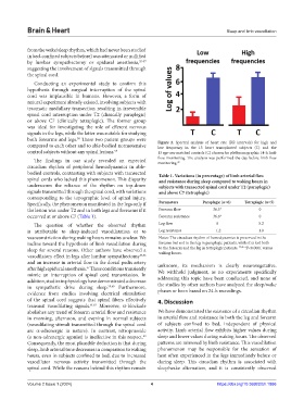
both forearms and legs. These two patient groups were Figure 2. Spectral analysis of heart rate (RR intervals) for high and
58
compared to each other and to able-bodied normotensive low frequency in the 13 heart transplanted subjects (T) and the
control subjects without any spinal lesions. 58 13 age-sex-matched controls (C) chosen for plethysmographic 14-h limb
The findings in our study revealed an expected flow monitoring. The analysis was performed the day before limb flow
monitoring.
49
circadian rhythm of peripheral hemodynamics in able-
bodied controls, contrasting with subjects with transected Table 1. Variations (in percentage) of limb arterial flow
spinal cords who lacked this phenomenon. This disparity and resistance during sleep compared to waking hours in
underscores the reliance of the rhythm on top-down subjects with transected spinal cord under T2 (paraplegic)
signals transmitted through the spinal cord, with variations and above C7 (tetraplegic)
corresponding to the topographic level of spinal injury.
Specifically, the phenomenon manifested in the legs only if Parameters Paraplegic (n=6) Tetraplegic (n=5)
the lesion was under T2 and in both legs and forearms if it Forearm flow 36.5* 0
occurred at or above C7 (Table 1). Forearm resistance 36.6* 0
The question of whether the observed rhythm Leg flow 0 −5.2
is attributable to sleep-induced vasodilatation or to Leg resistance −1.2 −1.0
vasoconstriction during waking hours remains unclear. We Notes: The circadian rhythm of hemodynamics is preserved in the
incline toward the hypothesis of limb vasodilation during forearm but not in the leg in paraplegic patients, while it is lost both
sleep for several reasons. Other authors have observed a to the forearm and the leg in tetraplegic patients. 17,49 *P<0.0001 versus
waking hours.
vasodilatory effect in legs after lumbar sympathectomy 55,56
and an increase in arterial flow in the dorsal pedis artery unknown, its mechanism is clearly neurovegetative.
57
after high epidural anesthesia. These conditions transiently We withhold judgment, as no experiments specifically
mimic an interruption of spinal cord transmission. In addressing this topic have been conducted, and none of
addition, studies in physiology have demonstrated a decrease
in sympathetic drive during sleep. 59,60 Furthermore, the studies by other authors have analyzed the sleep/wake
evidence from studies involving electrical stimulation phases or been based on 24-h recordings.
of the spinal cord suggests that spinal fibers effectively 4. Discussion
transmit vasodilating signals. 61,62 Moreover, α-blockade
abolishes any trend of forearm arterial flow and resistance We have demonstrated the existence of a circadian rhythm
in morning, afternoon, and evening in normal subjects in arterial flow and resistance in both the leg and forearm
(vasodilating stimuli transmitted through the spinal cord of subjects confined to bed, independent of physical
are α-adrenergic in nature). In contrast, nitroprusside activity. Limb arterial flow exhibits higher values during
(a non-adrenergic agonist) is ineffective in this respect. sleep and lower values during waking hours. The observed
63
Consequently, the most plausible deduction is that during patterns are mirrored by limb resistance. This vasodilation
sleep, limb arterial tone decreases in comparison to waking phenomenon may be responsible for the sensation of
hours, even in subjects confined to bed, due to increased heat often experienced in the legs immediately before or
vasodilator nervous activity transmitted through the during sleep. This circadian rhythm is associated with
spinal cord. While the reasons behind this rhythm remain sleep/wake alternation, and it is consistently observed
Volume 2 Issue 1 (2024) 4 https://doi.org/10.36922/bh.1886