
Page 52 - BH-3-3
P. 52
Brain & Heart A left atrial appendage occlusion review
Table 2. Summary of landmark and registry trials
Trial Conclusion
PROTECT-AF LAAC was both non-inferior and superior compared
to warfarin for the prevention of cardiovascular
death, stroke, or embolism, and was superior to
warfarin for all cause mortality
(95% CI: 0.41 – 1.05)
PREVAIL LAAC was non-inferior to warfarin for prevention
of ischemic stroke
(95% CI: −0.019 – 0.0273)
PRAGUE-17 LAAC was noninferior to DOAC in preventing
cardiovascular, neurological, and bleeding events
related to atrial fibrillation
ASAP LAAC with Watchman can be performed safely
without a transition period
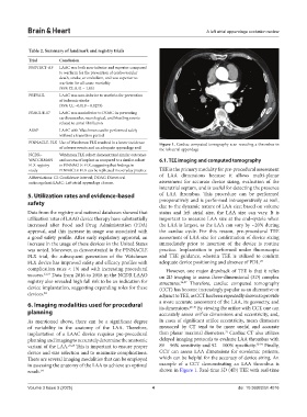
PINNACLE-FLX Use of Watchman FLX resulted in a lower incidence Figure 1. Cardiac computed tomography scan revealing a thrombus in
of adverse events and an adequate appendage seal the left atrial appendage
NCDR– Watchman FLX cohort demonstrated similar outcomes
WATCHMAN and success of implant as compared to a similar cohort 6.1. TEE imaging and computed tomography
FLX registry in PINNACLE-FLX, suggesting that findings in
study PINNACLE-FLX can be replicated in everyday practice TEE is the primary modality for pre-procedural assessment
Abbreviations: CI: Confidence interval; DOAC: Direct oral of LAA dimensions because it allows multi-planar
anticoagulant; LAAC: Left atrial appendage closure. assessment for accurate device sizing, evaluation of the
interatrial septum, and is useful for detecting the presence
5. Utilization rates and evidence-based of LAA thrombus. This procedure can be performed
safety preoperatively and is performed intraoperatively as well,
due to the dynamic nature of LAA size; based on volume
Data from the registry and national databases showed that status and left atrial size, the LAA size can vary. It is
utilization rates of LAAO device therapy have substantially important to measure LAA size at the end-systole when
increased after Food and Drug Administration (FDA) the LAA is largest, as the LAA can vary by ~20% during
approval, and this increase in usage was associated with the cardiac cycle. For this reason, pre-procedural TEE
a good safety profile. After early regulatory approval, an assessment of LAA size for confirmation of device sizing
increase in the usage of these devices in the United States immediately prior to insertion of the device is routine
was noted. Moreover, as demonstrated in the PINNACLE practice. Implantation is performed under fluoroscopic
FLX trial, the subsequent generation of the Watchman and TEE guidance, wherein TEE is utilized to confirm
FLX device has improved safety and efficacy profiles with adequate device positioning and absence of PDL. 23
complication rates < 1% and with increasing procedural However, one major drawback of TEE is that it relies
success. 22,23 Data from 2016 to 2018 in the NCDR LAAO on 2D imaging to assess three-dimensional (3D) complex
registry also revealed high fall risk to be an indication for structures. 26,27 Therefore, cardiac computed tomography
device implantation, suggesting expanding roles for these (CCT) has become increasingly popular as an alternative or
devices. 24 adjunct to TEE, as CCT has been repeatedly shown to provide
6. Imaging modalities used for procedural a more accurate assessment of the LAA, its geometry, and
By viewing the orifice with CCT, one can
its dimensions.
28-30
planning accurately assess orifice dimensions and eccentricity, and,
As mentioned above, there can be a significant degree in cases of significant orifice eccentricity, mean diameters
of variability in the anatomy of the LAA. Therefore, measured by CT tend to be more useful and accurate
31
implantation of a LAAC device requires pre-procedural than planar maximal diameters. Cardiac CT also utilizes
planning and imaging to accurately determine the anatomic delayed imaging protocols to evaluate LAA thrombus with
variant of the LAA. 23,25 This is important to ensure proper 89 – 96% sensitivity and 92 – 100% specificity. 32-34 Finally,
device and size selection and to minimize complications. CCT can assess LAA dimensions for euvolemic patients,
There are several imaging modalities that can be employed which can be helpful for the accuracy of device sizing. An
in assessing the anatomy of the LAA to achieve an optimal example of a CCT demonstrating an LAA thrombus is
result. 14 shown in Figure 1. Real-time 3D (4D) TEE with real-time
Volume 3 Issue 3 (2025) 4 doi: 10.36922/bh.4016