
Page 102 - GTM-3-1
P. 102
Global Translational Medicine Anergy in Leishmania-associated malignancy
Despite the inconclusive evidence, numerous studies
have linked Leishmania sp. to cancer. Pathologically,
6-8
the manifestations of leishmaniasis resemble those of
cancer, and their appearance may precede those of cancer.
A number of studies have reported the association between
Leishmania sp. and the development of malignant lesions
such as basal cell carcinoma, squamous cell carcinoma
(SCC), leukemia and lymphoma, and hemangiosarcoma.
6
Cutaneous SCC is the most common skin cancer among
blacks and the second most common among Whites,
Asians, and Hispanics. The mortality rate due to cutaneous
SCC ranges from 1.5% to 3.4%, with nodal metastases
occurring in 1.9% to 5.2% of cases, but early diagnosis can
7
improve the prognosis. The risk of developing cutaneous
SCC, the number of lesions, and the aggressiveness of
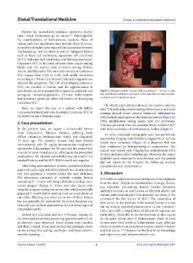
each lesion can be augmented by congenital, acquired, and Figure 1. Multiple nodular lesions, each measuring 5 – 12 mm in size,
iatrogenic immunosuppression. Chronic inflammation that were deeply infiltrated and fused to the skin with purulent exudate-
and infectious agents are other risk factors for developing oozing ulcers of irregular margins.
cutaneous SCC. 7
The Montenegro skin test showed non-reactive outcome
Here, we report the case of a patient with diffuse after 72 h, indicating positive anergy. Hematoxylin and eosin
cutaneous leishmaniasis who developed cutaneous SCC in staining showed severe chronic histiocytic inflammation
the active lesions of leishmaniasis. with multiple amastigotes in the tissue specimens (Figure 2).
2. Case presentation DNA amplification testing assays were not performed.
A biopsy procured from an ulcerated lesion unveiled SCC
In the present case, we report a 63-year-old farmer with mild coexistence of Leishmania sp. bodies (Figure 3).
from Comalcalco, Tabasco, Mexico, suffering from An axial computed tomography scan was performed,
diffuse cutaneous leishmaniasis, which was diagnosed uncovering imaging manifestations suggestive of cervical
10 years ago. The parasitic disease had been treated lymph node metastasis (Figure 4), a diagnosis that was
intermittently with 20 mg/kg intramuscular meglumine later confirmed by histopathological examination. The
antimonate (Glucantime) for 20 days, but the patient had patient was treated with 3 mg/kg/day amphotericin B for
a record of poor compliance in adhering to the prescribed 10 days and prescribed radiotherapy. However, the tumor
medications. No relevant comorbidity was reported in his exhibited poor response to radiotherapy, and the patient
medical history, and his HIV ELISA result was negative.
did not return to the hospital for follow-up medical
After being untreated for 6 months, several new lesions consultations and interventions.
appeared on the right side of the patient’s face, besides those
that first appeared 4 months before the new additions. 3. Discussion
The dermatosis consisted of multiple nodular lesions SCC is the second most common malignancy that originates
measuring 5 – 12 mm with deep infiltrates and deep, non- from the skin. Despite its multifactorial etiology, chronic
7
tender plaques (Figure 1). There were also ulcers with sun exposure, pre-existing lesions (actinic keratosis,
irregular margins oozing serous exudate, which reportedly unhealed wounds, or scar known as Marjolin ulcer), and
appeared 1 month before seeking medical consultation at chronic infections (mainly viral diseases) are some of the
our clinic. Bleeding was observed in two of these lesions prominent the risk factors of SCC. The occurrence of
7
but was generally left unattended. No actinic keratosis was skin cancer in the previous leishmaniasis lesions is a rare
detected, and cervical examination did not reveal signs of but an already reported phenomenon in the literature.
8
lymphadenopathy. Older men with a long history of ultraviolet exposure are
Several non-ulcerated and non-infiltrated nodules on particularly vulnerable to the development of skin cancer
the trunk and extremities measuring approximately 0.5 cm on the prior lesion sites of leishmaniasis. Most of these
in diameter were observed. These lesions were painless tumors arise from previous leishmaniasis scars, although
and flesh-colored. Some nodules had hemorrhagic crusts there are reports of an association between active infection
on the surface, fine scaling, and hyper- and hypo-chromic and skin cancer. 8-12 However, to the best of our knowledge
macular scarring. and experience, such an association is rare.
Volume 3 Issue 1 (2024) 2 https://doi.org/10.36922/gtm.2281