
Page 244 - IJB-10-1
P. 244
International Journal of Bioprinting NIR-secretome release for nerve regeneration
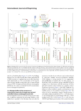
Figure 4. Biological effect on neural cells of the release of secretome through near-infrared radiation after induced damage. (A) Schematic representation
of the experimental setup. (B–D) Cell viability after the induction of damage with H O and controlled release of culture medium, astrocyte-conditioned
2
2
medium, and secretome of mesenchymal stem cells, respectively. (E–G) ROS production after the induction of damage with H O and controlled release
2
2
of culture medium, astrocyte-conditioned medium, and secretome of mesenchymal stem cells, respectively. Results are reported as % mean ± standard
deviation. **p < 0.05 and ***p < 0.01, one-way ANOVA and Tukey’s post-hoc test.
with the cell viability data (Figure 3A and B). Our findings secretome, but also the set of factors and vesicles released
indicate that for evaluating the effectiveness and potential by astrocytes (namely astrocyte-conditioned medium,
of MSCs secretome in regenerating neural cells, bioprinted ACM) displays a strong potential for the regeneration
AGO microbeads at a GO concentration of 0.5 mg/mL and proliferation of neural cells. For this purpose, we
offer the most suitable configuration due to their elevated bioprinted AGO microbeads having embedded ACM to
release of FITC–Dextran and their ability to downregulate compare their effectiveness with the secretome of MSCs
ROS levels. As a result, we selected this GO concentration and with standard culture medium as a negative control.
for our subsequent experiments. Results are reported in Figure 4. After NIR irradiation, the
release of culture medium caused a mild increase in cell
3.3. Biological effect of the secretome of viability, similar to the previous finding (Figure 4B). The
mesenchymal stem cells through near-infrared release of ACM, importantly, caused a strong increase in
controlled release on damaged neural cells cell viability, which resulted to be 67% ± 11.1% with respect
We then bioprinted AGO microbeads having GO at 0.5 mg/ to cells having induced neural damage with H O , which
2
2
mL, in which we embedded the secretome of MSCs. It has resulted to be 47% ± 9.3% (Figure 4C, p < 0.05, one-way
widely been reported in the literature that not only the ANOVA and Tukey’s post-hoc test). After controlled release
Volume 10 Issue 1 (2024) 236 https://doi.org/10.36922/ijb.1045