
Page 423 - IJB-10-2
P. 423
International Journal of Bioprinting In vitro 3D pancreatic acinar unit
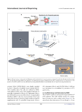
Figure 1. 3D layer-by-layer in vitro model. (a) Illustration of the functional unit of exocrine pancreas, composed by epithelial cells surrounded by stromal
cells. (b) Melt-electrowriting (MEW) was employed: CAD drawings, processing of polycaprolactone (PCL), and production of a microscopic in vitro
model. (c) The MEW scaffolds were then cellularized by seeding human fibroblasts (i) and epithelial cells (ii). Figure drawn using Biorender.com.
activated KRAS (HPDE-KRAS)—were kindly provided 2% L-glutamine (Gibco), and 15% FBS (Gibco). Cell lines
by Prof. F. Bussolino (Candiolo Cancer Institute-IRCCS- were maintained in a humidified CO incubator at 37°C
2
FPO, Candiolo, Italy). The cells were cultured in RPMI- and 5% CO .
1640 medium (Gibco, Jenks, USA) supplemented with 1% 2
penicillin–streptomycin (Gibco), 1% L-glutamine (Gibco), 2.2. Scaffold design and fabrication by MEW
®
and 10% fetal bovine serum (FBS; Gibco). Human foreskin The 3D MEW models were designed through SolidWorks
fibroblasts (HFF1) cells were obtained from ATCC and CAD software. The CAD models consist of a square-
®
cultured in Dulbecco’s Modified Eagle’s Medium (DMEM) based 3D structure with a central cavity (Figure 1c). The
supplemented with 1% penicillin–streptomycin (Gibco), cuboid had a final dimension of 10 mm (length) × 10 mm
Volume 10 Issue 2 (2024) 415 doi: 10.36922/ijb.1975