
Page 12 - OR-1-2
P. 12
Parkinson’s research, focusing on their role in elucidating by DA neuron loss and increased apoptosis, mirroring
disease pathomechanisms (Table 1). We then explore neurodegenerative processes observed in PD brains. 20-22
their applications in drug discovery and high-throughput Single-cell RNA sequencing of SNCA triplication hMOs has
screening. Finally, we discuss the potential of organoid provided insights into molecular dysfunctions affecting DA
transplantation as a therapeutic strategy for PD. neurons, including impaired oxidative phosphorylation,
dysregulated protein translation, and endoplasmic
2. Organoid models for PD reticulum stress. Furthermore, recent studies suggest that
23
2.1. SNCA α-synuclein pathology is linked to cellular senescence,
particularly in astrocytes, leading to a phenomenon termed
α-Synuclein, a 140-amino-acid presynaptic neuronal astrosenescence, supporting that pathological α-synuclein
protein encoded by the SNCA gene, plays a critical role may contribute to the induction of neuroinflammation,
in synaptic function, but its pathological aggregation which ultimately increases the susceptibility of DA neurons
into Lewy bodies is a hallmark of PD. SNCA mutations, to degeneration. 22
18
particularly missense mutations and gene amplifications,
are associated with autosomal dominant PD. Among In addition to using hMOs to investigate the potential
these, SNCA triplications lead to a substantial increase in cellular mechanisms of SNCA-induced neuronal toxicity,
α-synuclein expression, accelerating pathological processes organoid models have also been employed to study the
and neurodegeneration. Studies using hMOs derived propagation of α-synuclein pathology. In a recent study,
19
from iPSCs carrying SNCA triplication mutations have researchers used mouse intestinal organoids expressing
successfully replicated key features of α-synuclein pathology human α-synuclein to examine the transfer of α-synuclein
seen in patients with synucleinopathies. hMOs with SNCA from epithelial cells within the organoids to co-cultured
triplications exhibit elevated α-synuclein levels and a time- nodose neurons lacking α-synuclein expression. This
dependent increase in α-synuclein aggregation. 20-22 This study highlights a potential non-neuronal source of
aggregation includes both oligomeric and phosphorylated fibrillar α-synuclein, suggesting that gut mucosal cells
forms of α-synuclein, which are detected in both may contribute to the initiation or spread of α-synuclein
neurons and glial cells. The progressive accumulation of pathology. In addition to the genetic manipulation of
24
20
pathological α-synuclein in these organoids is accompanied SNCA, hMOs are also utilized to study the propagation of
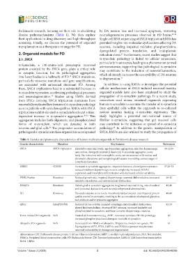
Table 1. Genetic and phenotypic characteristics of human midbrain organoids in Parkinson’s disease research
Genetic characteristic Mutation Key features References
SNCA SNCA triplication Elevated α-synuclein levels, age-dependent aggregation, selective dopaminergic 20-22,56
neuron loss, detergent-resistant β-sheet-rich α-synuclein aggregates, Lewy
body-like inclusions with eosinophilic cores, astrosenescence with nuclear and
chromatin alterations, and morphological features resembling various stages of
Lewy body formation.
LRRK2 G2019S Increased α-synuclein aggregation, impaired clearance, altered gene expression, 17,28-31
reduced midbrain dopaminergic neuron complexity, increased FOXA2
expression, and incomplete differentiation with decreased cellular variability.
PINK1/Parkin Knockout Reduced growth rate, impaired dopaminergic neuronal differentiation, increased 38-42
oxidative stress levels, and mitochondrial dysfunction.
DNAJC6 Knockout Pathological α-synuclein aggregation, heightened neuronal firing, mitochondrial 45,46
and lysosomal dysfunction, and neurodevelopmental abnormalities.
DJ1 Knockout Elevated oxidative stress levels, mitochondrial dysfunction, and impaired protein 48,49
quality control in astrocytes, resulting in the accumulation of advanced glycation
end products and α-synuclein aggregates
GBA1 L444P/N370S Reduced GCase activity, impaired autophagy, mitochondrial dysfunction, 52-59
altered lipid metabolism, decreased TH neurons, increased insoluble and
+
phosphorylated α-synuclein, and fewer complex dopaminergic neurons.
Toxin-induced PD organoids N/A Treated with neurotoxins (e.g., MPP , rotenone) to induce PD-like pathology. 60
+
Increased phosphorylated and detergent-insoluble α-synuclein.
Idiopathic PD organoids N/A Generated from PBMCs of idiopathic PD patients, models non-genetic PD. 61
Dysregulation of TH, PTX3, LMX1A, and FOXA2 expression may increase
neuronal vulnerability to damage and degeneration
Abbreviations: DJI: Parkinson’s disease protein 7; GCase: Glucocerebrosidase; MPP : 1-methyl-4-phenylpyridinium; N/A: Not available;
+
PBMCs: Peripheral blood mononuclear cells; PD: Parkinson’s disease; TH: Tyrosine hydroxylase; LMX1A: LIM homeobox transcription
factor 1 alpha.
Volume 1 Issue 2 (2025) 3 doi: 10.36922/OR025040006