
Page 90 - AN-1-3
P. 90
Advanced Neurology A case report of epileptic vertigo
A B
C D
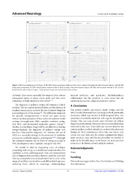
Figure 1. Electroencephalogram findings. (A–B) EEG shows paroxysmal spike and slow wave complex discharges in both temporal regions, with the left
being more prominent. (C) EEG shows phase reversal in the T1 lead, located at the left temporal region. (D) EEG shows phase reversal in the T2 lead,
located at the right temporal region. These epileptic waves are marked by the blue arrows.
the brain. These areas, especially the temporal lobe, release internal medicine, and psychiatry. Multidisciplinary
paroxysmal spikes or sharp waves, spike and slow wave collaboration has the potential to cover areas that are
complexes, or high amplitude slow waves [1,11] . underemphasized in a single professional context.
The diagnosis of epileptic vertigo still remains a clinical 4. Conclusion
problem. The incomplete medical history and the absence of
ancillary examinations increase the rate of missed diagnosis This patient initially experienced simple vertigo, and her
and misdiagnosis of this disease . The differential diagnosis EEG revealed abnormal wave discharges at both sphenoidal
[12]
for episodic vertigo/dizziness is broad and spans across electrodes, which were located at both temporal lobes. Her
various medical specialties. In fact, only a few patients would symptoms remarkedly improved with regular antiepileptic
undergo thorough brain MRI, complete vestibular testing, therapy. This case may provide some reference for clinical
ictal EEG, and professional molecular genetic testing . diagnosis and treatment. However, there are some limitations
[13]
Besides, if antiepileptic drugs (AEDs) are effective in treating observed in this case. Video EEG test was not done for the
vertigo/dizziness, the diagnosis of epileptic vertigo may patient, neither was flash stimulation or sleep induction used
also be a false-positive diagnosis. For instance, the use of during her EEG examination. Since this case report only
AEDs is a successful strategy for the treatment of vestibular covers one case, there may be certain contingencies; hence,
paroxysmia, vestibular migraine, and migraine [14,15] . Although more practical cases and theoretical studies are needed to
the treatment response rate of AEDs in vertigo is as high as make up for the deficiency. In the future, we must pay close
90%, the diagnostic rate of epileptic vertigo is only 10%. attention to the history of such epileptic patients.
We should be alert in diagnosing cases of epilepsy Acknowledgments
presenting with vertigo as a standalone symptom since this
condition is often underdiagnosed. This case demonstrates None.
that to diagnose this condition, there is a need for a detailed
history, a comprehensive physical examination, and a wide Funding
range of ancillary examinations and differential diagnoses, This work was supported by Yuan Du Scholars and Weifang
including those related to neurology, otolaryngology, Key Laboratory.
Volume 1 Issue 3 (2022) 3 https://doi.org/10.36922/an.v1i3.140