
Page 10 - AN-2-2
P. 10
Advanced Neurology Venous stenting, intracranial hypertension
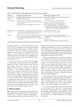
Table 1. Comparison of procedural alternatives to cerebral venous sinus stenting
Procedure Advantages (compared to CVSS) Disadvantages (compared to CVSS)
Repeated lumbar (i) Slight drop in IIH and headache immediately post-procedure [45] (i) Rebound IIH with longer and more severe headache than
punctures (ii) Normalizes CSF pressure [19] before the procedure [45]
Bariatric surgery (i) Significant reduction in headaches (90% reduction with (i) Can only be used for IIH patients with obesity
bariatric surgery versus 70.2% reduction with CVSS), more (ii) More pre-operative and post-operative care
than in CVSS [21,22] (iii) Variable cost but more expensive on average [42,43]
(ii)Comparable reductions in papilledema and tinnitus [21,41] (iv) Very little improvement in visual complaints [41]
(v) Normally offered after failure of weight management
interventions, thereby requiring a longer time before any
improvement of IIH symptoms can be observed [43]
Optic nerve sheath (i) Highly effective at treating visual impairment symptoms of (i) Does not reduce headaches in IIH [47]
fenestration IIH (such as decreasing papilledema grade and increasing (ii) Less improvement in visual acuity [44]
visual field measured through kinetic perimetry) [47]
Shunting (i) Better improvement in visual acuity than in repeated lumbar (i) Less improvement in visual acuity and headaches [21]
punctures, but not as significant as in ONSF or CVSS [44] (ii) Moderate need for repeat procedures, making it more costly
(ii) Reduction in postoperative headaches, but not as significant overall [21]
as in CVSS [44] (iii) More invasive [21]
(iv) Significant complication rate as compared to CVSS [21]
CVSS: Cerebral venous sinus stenting; IIH: Idiopathic intracranial hypertension.
the general population, in which the incidence was only CVSS has been chosen as the course of action, aspirin and
6.8% . By current standards, dural venous sinus stenosis clopidogrel should be taken 3–4 days before stent placement
[4]
can be described as extrinsic, intrinsic, or both . Extrinsic and continued for 3–6 months after which only aspirin is
[26]
[21]
stenosis can be related to scarring or elevated intracranial used . Although oral anticoagulants with single antiplatelet
hypertension and presents as a long, smooth narrowing, therapy may be better in preventing intra-stent thrombosis,
whereas an intrinsic obstruction pattern consists of an dual antiplatelet therapy has been documented in literature
[27]
arachnoid granulation defect that causes focal filling of the as being more frequently used in CVSS .
vein, which presents as a round or oval-shaped formation Venous sinus stenting is done under general anesthesia,
in the dural sinus wall . and a heparin bolus is given as soon as venous access
[25]
[4]
The first venous stent was placed in 2002 by Higgens is achieved to prevent clotting . Although most stent
et al. In the study, a catheter was inserted into the internal surgeries use the femoral vein as an access site, upper
jugular vein to direct a stent into the stenosed transverse extremity access through the brachial or basilic vein can
[28]
sinus, which resulted in a decrease in the pressure also be used in CVSS . Either the right or left internal
[28]
gradient from 18 mmHg pre-procedure to 3 mmHg post- jugular vein can be accessed using a right-arm approach .
procedure [8,24] . The researchers described a significant Single arm access allows for earlier patient mobility,
improvement in symptoms after the placement of stent. In easier monitoring of healing, shorter procedure time, and
a meta-analysis by Mufti et al., 367 cases from 25 different reduction of femoral vein access site complications, such as
publications were analyzed. Of these patients receiving one retroperitoneal hemorrhage [26,28] .
or more venous sinus stents, 78% experienced improvement A microwire is guided through a micro catheter into the
in visual acuity and 77% had headache resolution; among superior sagittal sinus and then past the point of stenosis .
[4]
the patients with papilledema, 84.5% showed resolution Maximum venous pressures are reconfirmed at various
or improvement; and among the patients with pulsatile spots in the dural venous sinus system (internal jugular,
tinnitus, 88.7% had resolution after stent placement. CVSS jugular bulb, bilateral transverse sinus, superior sagittal
has been shown to have high technical success, low relapse sinus, and sigmoid sinus) using venous manometry .
[4]
rates, and low major overall complication rates . The access sheath system for the stent normally consists
[26]
5. CVSS procedure of three sheaths (7F, 9F, and 12F) of varying lengths
(80–90 cm) . Although most CVSS is performed using
[4]
Before stenting, patients need to be screened for papilledema, self-expanding stents, balloon-expanding stents may be
their baseline visual acuity should be noted, and appropriate considered as well . Stents are placed across the stenosis,
[26]
imaging such as magnetic resonance angiography or extending from 10 mm before the stenosis to 10 mm after
venography with manometry should be performed . Once the stenosis .
[4]
[4]
Volume 2 Issue 2 (2023) 4 https://doi.org/10.36922/an.284