
Page 57 - AN-3-1
P. 57
Advanced Neurology Dementia with Lewy bodies and substance misuse
hyponatremia, possibly trazodone-related SIADH. Renal
29
and hepatic 30,31 panels were normal, with no signs of peripheral
organ disease. Magnetic resonance angiography, magnetic
resonance venography, magnetic resonance imaging (MRI)
with and without contrast, and computed tomography (CT)
of the brain in May 2016 showed no abnormal findings. 32,33
Repeated MRI and CT of the brain in February 2017 revealed
mild small vessel ischemic disease and cerebral atrophy with
chronic white matter microvascular ischemic disease, a
finding believed to be linked to normal aging. 34
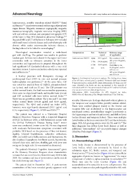
Neurological examination revealed a wide-based Interpretation Test Technical Reference range
gait with shuffling. The patient was unable to perform report (if applicable)
tandem gait. Reflexes were absent in the upper and lower Indeterminate-NADD Aβ42 293.1 pg/mL Not consistent with
extremities with no vibratory sensation in the lower T-Tau 136.26 pg/mL AD: p-tau<54 pg/mL,
extremities and hypoesthesia to pinprick throughout. He p-Tau 32.65 pg/mL ATI>1.2
had significant left dysdiadochokinesia with a very mild ATI 0.74 Borderline: p-tau
54-68 pg/mL and/or
intention tremor. A nerve conduction velocity test reported ATI 0.8–1.2
normal latencies. 35 AD: p-tau>68 pg/mL
and ATI<0.8
A lumbar puncture with therapeutic drainage of
cerebrospinal fluid (CSF) to rule out normal pressure Figure 1. Cerebrospinal fluid protein analysis. The finding above shows
36
hydrocephalus was performed. At the same time, CSF a low ATI but a normal p-tau (+ symbol): “Reduced Aβ42 and elevated
T-tau levels lead to a reduced ATI, which differentiates AD from non-AD
was tested for amyloid-beta 42 (Aβ42), phosphorylated- cases. Elevated p-tau levels are also encountered in the majority of
tau (p-tau), and total-tau (T-tau). The CSF pressure was individuals with AD. The current result of reduced ATI and normal levels
within normal limits, the fluid was normal in appearance, of p-tau are of unknown clinical significance.
there were no oligoclonal bands, and myelin basic protein Abbreviations: Aβ42: Amyloid-beta 42; ATI: Amyloid tau index;
and CSF nucleated cells were within normal limits. 37-39 NADD: Non-AD dementia; p-Tau: Phosphorylated-tau; T-Tau: Total-tau.
There was no growth in culture. The p-tau and T-tau were atrophy. Microscopic findings displayed subpial gliosis in
within normal limits (136.26 pg/mL and 32.65 pg/mL, the temporal and occipital lobes, possibly seizure related.
respectively). The Aβ42 and amyloid tau index (ATI), There were amyloid plaques (rarely in the frontal and
however, were significantly decreased (293.1 pg/mL and
0.74, respectively) (Figure 1). APOE was Ɛ3/Ɛ4. 40-43 occipital lobe and moderately in the hippocampus and
entorhinal cortex), neurofibrillary tangles (moderately in
The patient was referred to a University Hospital the hippocampus and entorhinal cortex), and diffuse Lewy
Memory Disorders Program with a suspected diagnosis bodies (frontal and temporal lobes). There were multiple
of DLB. In February 2018, a full Parkinson’s screen with Lewy bodies in the locus coeruleus (LC) and the substantia
the Unified Parkinson’s Disease Rating Scale motor nigra. NIA-AA grading criteria were A2, B2, and C2. The
44
46
score was conducted (motor score 34). He was diagnosed final diagnosis was diffuse Lewy body disease as well as
with moderate Parkinsonism symptoms consistent with additional “intermediate” AD neuropathological changes
probable DLB based on the presence of fine rest tremor, (MedStar Pathology) (Figure 3).
rigidity, bilateral bradykinesia, subjective orthostasis,
RBD, and mild tactile hallucinations and fluctuations. He 3. Discussion
also displayed hypophonic but fluent speech, hypomimia, 3.1. Neuropathology of DLB
delayed chair raise, and slow, shuffling gait with poor arm
swing on the right side. He was started on donepezil. Lewy body disease is characterized by the presence of
Lewy bodies, which can commonly be found in the
The patient’s Montreal Cognitive Assessments given substantia nigra, LC, limbic system, and temporal and
45
at the Memory Disorders Program show characteristic frontal cortex. 47,48 Lewy bodies (Figure 4A) are intracellular
variance but an overall decline from February 2018 to July aggregates of proteins, known as “aggresomes,” the major
2019. The variance and loss of perseverance can be seen in component of which is alpha-synuclein (α-synuclein). 49,50
his clock drawing task (Figure 2). There may also be Lewy neurites (Figure 4B) and
The patient died in December 2021. A post-mortem accumulations of α-synuclein in nerve cell processes. The
examination of his brain revealed grossly apparent mild majority of cases are also pathologically related to AD, with
Volume 3 Issue 1 (2024) 5 https://doi.org/10.36922/an.2232