
Page 12 - AN-3-2
P. 12
Advanced Neurology Limbic-predominant TDP-43 encephalopathy
compared to remote memory) and slow and progressive clinical data suggesting the existence of synucleinopathy).
5
involvement of other cognitive domains in dementia There are also non-mandatory supportive criteria,
stages. 5,8,26,46 Therefore, it is not surprising that LATE is including: (i) clinical factors (patient age equal to or greater
part of the potential differential diagnosis of AD. 11,29,39 than 75 years; relatively preserved neocortical functions;
However, it is worth noting that pure cases of LATE affect the presence of semantic memory impairment in early
individuals of more advanced age compared to pure cases stages of the disease); (ii) structural (disproportionate
of AD or cases with co-pathology of AD and LATE. In hippocampal atrophy in MRI) and functional
addition, LATE cases have a more benign clinical course neuroimaging criteria (FDG-PET [fluorodeoxyglucose-
with a slower progression of amnestic cognitive decline. positron emission tomography] hypometabolism in the
9
Therefore, the diagnosis of possible LATE should be limbic system without a characteristic pattern of AD); and
considered, to be confirmed through anatomopathological (iii) low likelihood of significant neocortical tau pathology
study, in individuals with a typical clinical syndrome of (confirmation of low probability of AD pathology assessed
AD (predominantly amnestic MCI), a negative core AD through ATN biomarkers in CSF or amyloid PET). The
52
biomarker study, advanced age, and in the absence of degree of diagnostic certainty is established based on
another potential alternative etiological diagnosis. the number of supportive criteria met in addition to the
The main challenge in achieving an early and accurate mandatory criterion of amnestic-predominant cognitive
diagnosis of LATE lies in the absence of validated clinical decline.
criteria for use in clinical practice and the lack of reliable In addition to the predominantly amnestic cognitive
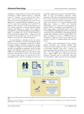
biomarkers (Figure 2). Recently, a proposal has emerged decline, researchers have explored whether other
5
to establish the diagnosis of LANS (limbic-predominant behavioral or motor symptoms are more or less prevalent
amnestic neurodegenerative syndrome), which would also in individuals diagnosed with LATE. However, the
13
suggest LATE, although it would not be specific to it. The results regarding behavioral symptoms are inconsistent to
52
diagnosis of LANS requires the presence of insidious, slowly date. 34,35 One study suggests that cases of pure LATE-NC
progressive cognitive decline over 2 or more years, with a have a slightly higher risk of developing behavioral
clear amnestic predominance, and the absence of other symptoms associated with the frontal lobe, while pure
identifiable causes to justify it (including the absence of AD cases exhibit a mild increase in the risk of agitation;
Figure 2. Typical clinical features of a patient with possible pure limbic-predominant age-related TDP-43 encephalopathy. Image created using Biorender.
com
Abbreviations: AT(N): Amyloid, tau, and neurodegeneration; CSF: Cerebrospinal fluid; FDG-PET: Fluorodeoxyglucose-positron emission tomography;
MRI: Magnetic resonance imaging.
Volume 3 Issue 2 (2024) 6 doi: 10.36922/an.2603