
Page 110 - AN-3-4
P. 110
Advanced Neurology Stress accelerates parkinsonism in rats
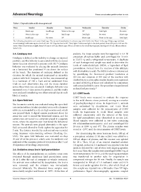
Table 1. Unpredictable mild stress protocol
Week Sunday Monday Tuesday Wednesday Thursday Friday
1 Moist cage Small cage Water in the cage 30° Total light No water
2 Water in the cage 30° Small cage Total light No water Moist cage
3 Small cage No water 30° Moist cage Water in the cage Total light
The following procedures were used: Small cage: The rat was housed alone in a mouse cage (29×18×12 cm). Water in the cage: Empty cage (without
wood shavings) with water up to a level of 2 cm. 30°: Cage inclined at 30°. Total light: Continuous light in the dark period, from 6:30 pm to 6:30 am. No
water: Water deprivation at night, from 6:30 pm to 6:30 am. Moist cage: 250 mL of water in the wood shaving layer, starting at 6:30 am. All procedures
lasted 12 h.
2.4. Catalepsy test analysis, the tissue samples were homogenized in 0.1 M
potassium phosphate buffer and centrifuged for 10 min
Catalepsy is defined as the inability to change an imposed
position, and this behavior is associated with the decreased at 22,673 ×g and a refrigerated temperature. A duplicate
motor function observed in patients with PD. Catalepsy of each homogenized sample was used to determine the
38
behavior was evaluated by placing the animal’s forepaws levels of malondialdehyde (MDA, a byproduct of lipid
on a horizontal bar positioned 9 cm above the surface peroxidation, formed from the reaction of this aldehyde
of the bench. The duration of catalepsy, defined as the with thiobarbituric acid). The MDA levels were determined
duration for which the animal maintained an immobile by quantifying the fluorescent product (excitation at
posture with both forepaws on the bar, was measured up 315 nm and emission at 553 nm) of the reaction with
to a maximum of 180 s. Each animal underwent three thiobarbituric acid in a plate reader. Results were expressed
trials on each observation day, and the mean duration as nmol of MDA/g of tissue and calculated by comparison
across these trials was calculated. Cataleptic behavior was with a standard MDA curve. The procedure was performed
29
measured every 3 days across the protocol, and the results as described previously.
were analyzed considering two observational days in each 2.7. CORT levels
block of results.
CORT levels were measured to evaluate the response
2.5. Open field test to the mild chronic stress protocol, serving as a marker
The locomotor activity was evaluated using the open field of psychophysiological stress. In Experiment I, animals
test conducted in a circular open field arena with a diameter were euthanized by decapitation, and trunk blood
of 84 cm, surrounded by a 32-cm high wooden wall, which samples were collected for the measurement of CORT
was painted black. A digital camera positioned above the levels. In Experiment II, trunk blood samples were
arena was used to record the behavioral session, and the collected concurrently with the removal of the brain
camera was connected to a computer placed in a separate for lipid peroxidation assay (described in section 2.6).
room, where the experimenter monitored the behavioral Blood samples were collected in test tubes containing
session. The animals were placed alternately in the open 6% ethylenediaminetetraacetic acid and centrifuged for
field arena and allowed to freely explore the apparatus for 20 min at 1209 ×g and 4°C. The plasma samples were
5 min. The distance traveled by each animal was recorded stored at −20°C until the determination of CORT levels.
using Anymaze video-tracking software (Stoelting Co., For determining the stress hormone levels, 100 μL of
USA). The open field behavior was evaluated on days a precipitant solution (1 M zinc sulfate) was added to a
19 and 29 of the protocol, which were the midpoint and 100-μL aliquot of plasma, and the mixture was agitated
endpoint of the reserpine protocol (evaluations after 5 and for 30 s. Then, 50 μL of an internal standard solution
10 injections of the drug), respectively. (10 μg/mL cortisol in 1:1 methanol) was pipetted into this
mixture, followed by 1 mL of ether, with ongoing agitation
2.6. Oxidative stress: brain lipid peroxidation for 1 min. Next, the samples were centrifuged (1086.5 ×g, at
The effects of the manipulations on oxidative stress were room temperature) for 5 min, and the resulting supernatant
evaluated through membrane lipid peroxidation assay. was transferred to another tube for evaporation in
At 48 h after the end of reserpine or vehicle treatment, compressed nitrogen for 30 min. Finally, the sample was
the animals were euthanized by decapitation, the brains resuspended in 100 μL of 1:1 methanol: water and 0.1%
were removed, and the striatum was immediately formic acid under agitation for 10 s, after which 20 μL of
dissected bilaterally. The dissected tissue samples were this sample was injected into a Shimadzu Class VP high-
immediately weighed and frozen at −80°C. At the time of performance liquid chromatography apparatus coupled
Volume 3 Issue 4 (2024) 4 doi: 10.36922/an.4037