
Page 33 - AN-4-1
P. 33
Advanced Neurology Ferroptosis in neonatal HIBI
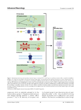
Figure 1. Mechanisms of primary and secondary neuronal injury in neonatal HIBI. Under hypoxic conditions, neurons rely on anaerobic metabolism,
leading to ATP depletion and acidosis in the primary neuronal injury phase. Another primary neuronal injury mechanism is anoxic depolarization. This
results in dysregulated ion and water accumulation, promoting free radical production and cell swelling. Mitochondrial dysfunction also contributes by
reducing ATP production and stimulating free radical release. Excessive glutamate accumulation leads to NMDA receptor hyperactivation, promoting
calcium-dependent free radical production. Microglia adopt phagocytic properties and release proinflammatory cytokines and chemokines, facilitating
the recruitment of peripheral immune cells into the brain. Effector molecules from these primary neuronal injury mechanisms promote larger-scale
secondary cell death through apoptosis, necrosis, autophagy, and ferroptosis, each with its distinct mechanisms and cellular features. Figure was created
using BioRender.com.
Abbreviations: HIBI: Hypoxic-ischemic brain injury; NMDA: N-methyl-D-aspartate.
compounds, which are selectively activated by the Ras the Stockwell group’s former observations that cell death
family members, a group of >150 guanosine triphosphatases induced by erastin – an RSL – does not exhibit the defining
with essential signaling functions in diverse cellular features of apoptosis and is unaffected by a pan-caspase
processes. 31,32 Key to the discovery of ferroptosis were inhibitor. They therefore concluded that it occurs through
Volume 4 Issue 1 (2025) 27 doi: 10.36922/an.4575