
Page 135 - AN-4-2
P. 135
Advanced Neurology Management of subdural hygroma and intracranial ACs
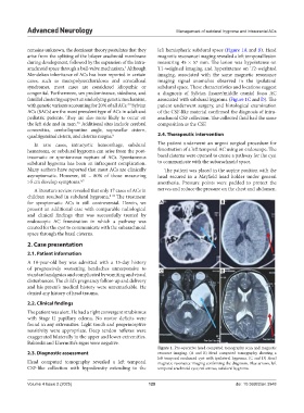
remains unknown, the dominant theory postulates that they left hemispheric subdural space (Figure 1A and B). Head
arise from the splitting of the bilayer arachnoid membrane magnetic resonance imaging revealed a left temporal lesion
during development, followed by the expansion of the intra- measuring 45 × 57 mm. The lesion was hypointense on
arachnoid space through a ball-valve mechanism. Although T1-weighted imaging and hyperintense on T2-weighted
2
Mendelian inheritance of ACs has been reported in certain imaging, associated with the same magnetic resonance
cases, such as mucopolysaccharidoses and acrocallosal imaging signal anomalies observed in the ipsilateral
syndromes, most cases are considered idiopathic or subdural space. These characteristics and locations suggest
congenital. Furthermore, sex predominance, sidedness, and a diagnosis of Sylvian fissure/middle cranial fossa AC
familial clustering support an underlying genetic mechanism, associated with subdural hygroma (Figure 1C and D). The
with genetic variants accounting for 20% of all ACs. Sylvian patient underwent surgery, and histological examination
2,3
ACs (SACs) are the most prevalent type of ACs in adult and of the CSF-like material confirmed the diagnosis of intra-
pediatric patients. They are also more likely to occur on arachnoid CSF collection. The collected fluid had the same
the left side and in men. Additional sites include cerebral composition as the CSF.
4,5
convexities, cerebellopontine angle, suprasellar cistern,
quadrigeminal cistern, and cisterna magna. 6 2.4. Therapeutic intervention
In rare cases, intracystic hemorrhage, subdural The patient underwent an urgent surgical procedure for
hematoma, or subdural hygroma can arise from the post- fenestration of a left temporal AC using an endoscope. The
traumatic or spontaneous rupture of ACs. Spontaneous basal cisterns were opened to create a pathway for the cyst
subdural hygroma has been an infrequent complication. to communicate with the subarachnoid space.
Many authors have reported that most ACs are clinically The patient was placed in the supine position with the
asymptomatic. However, 60 – 80% of those measuring head secured in a Mayfield head holder under general
>5 cm develop symptoms. 6,7 anesthesia. Pressure points were padded to protect the
A literature review revealed that only 17 cases of ACs in nerves and reduce the pressure on the chest and abdomen.
children resulted in subdural hygroma. 8-24 The treatment
for symptomatic ACs is still controversial. Herein, we A B
present an additional case with comparable radiological
and clinical findings that was successfully treated by
endoscopic AC fenestration in which a pathway was
created for the cyst to communicate with the subarachnoid
space through the basal cisterns.
2. Case presentation
2.1. Patient information
A 10-year-old boy was admitted with a 15-day history
of progressively worsening headaches unresponsive to C D
standard analgesics and complicated by vomiting and visual
disturbances. The child’s pregnancy follow-up and delivery
and his parent’s medical history were unremarkable. He
denied any history of head trauma.
2.2. Clinical findings
The patient was alert. He had a right convergent strabismus
with Stage II papillary edema. No motor deficits were
found in any extremities. Light touch and proprioceptive
sensitivity were appropriate. Deep tendon reflexes were
exaggerated bilaterally in the upper and lower extremities.
Babinski and Lhermitte’s signs were negative.
Figure 1. Pre-operative head computed tomography scan and magnetic
2.3. Diagnostic assessment resource imaging. (A and B) Head computed tomography showing a
left temporal arachnoid cyst with ipsilateral hygroma. (C and D) Head
Head computed tomography revealed a left temporal magnetic resonance imaging confirming the diagnosis. Blue arrows, left
CSF-like collection with hypodensity extending to the temporal arachnoid cyst; red arrows, subdural hygroma.
Volume 4 Issue 2 (2025) 129 doi: 10.36922/an.3948