
Page 136 - AN-4-2
P. 136
Advanced Neurology Management of subdural hygroma and intracranial ACs
The head was turned away from the surgeon, allowing for 2.5.1. Summary of the key points (Table 1)
a 90-degree turn. An incision was made above the zygoma, Summary of the treatment and outcome of a temporal
behind the hairline. A 1.5 cm incision was made through AC complicated with subdural hygroma in the pediatric
the temporalis muscle to the underlying temporal bone. population is shown in Table 1. Middle fossa AC cysts can
A small burr hole was created near the inferior aspect experience the following:
of the incision. Through the dura, which was cruciately • Spontaneous enlargement, followed by its disappearance
incised and sutured outward, a dark blue tinge was without clinical symptoms.
observed. The CSF freely exited from the wound using • Spontaneous enlargement and post-traumatic rupture
pressure, and the endoscope was introduced. At this point, resulting in subdural hematoma or hygroma. They are
landmark identification was crucial, and the temporal floor typically asymptomatic.
and tentorium served as the main landmarks. The internal • Spontaneous enlargement and spontaneous rupture
carotid artery and second and third cranial nerves should resulting in a subdural hematoma or hygroma. They
be identified. The arachnoid tissue, which was draped are typically symptomatic.
across and between the structures, was notably thicker and • The treatment for symptomatic ACs is still controversial.
grossly abnormal, resembling billowing sheer curtains. • Endoscopic AC fenestration to create a pathway for
These arachnoid layers were sliced to reveal the posterior the cyst to communicate with the subarachnoid space
communicating artery and internal carotid artery and through the basal cisterns is now possible with good
permit CSF circulation. Before locating and opening outcomes in experienced hands.
the thickened membrane of Lillequist, the posterior
communicating artery, and third nerve were completely 3. Discussion
fenestrated by the thickened arachnoid. The basilar artery,
which was located directly below the thicker arachnoid Although middle fossa ACs rarely undergo spontaneous
segment, and all other perforating vessels needed careful enlargement, disappearance, or rupture that results in
protection. After the fenestration was made, a broad a subdural hematoma or hygroma, they are typically
25
passageway leading into the basilar cisterns was constructed asymptomatic. In adults, the AC size usually increases by
to facilitate the observation of the contralateral third nerve, 2 – 3%, which is less than that of the pediatric population.
26
posterior cerebral artery, superior cerebellar artery, and Headache is the most typical sign of middle fossa ACs.
basilar artery with its perforators. At this time, good CSF Interestingly, most complications have been associated
flow was apparent. After the usual closure, the dura was with middle cranial fossa cysts, and all cases of cyst rupture
closed, and the bone powder was used to seal the burr hole. resulting in subdural hygroma have been reported in the
context of mild head injuries. 27,28 These findings could be
2.5. Follow-up explained by various factors, such as the disappearance of
Postoperatively, the patient was doing well and still alert. cysts following severe trauma or death of the patient from
He had no motor–sensory deficits. The intracranial trauma, malignant complications such as severe subdural
hypertensive syndrome subsided. One month after surgery, or intracystic hemorrhages concealing a prior hygroma, or
the boy was in good health with satisfactory post-operative failure to notice cysts in the presence of multiple injuries. 29
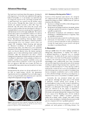
computed tomography findings (Figure 2). Conversely, the case presented refuted any prior
head injuries. Although the exact cause of the rupture
is unknown, theories include direct damage to the
ipsilateral sphenoid wing from the thinned temporal bone
or compression of the Sylvian ACs against it. Cyst wall
disruption and subsequent CSF leakage into the subdural
space are believed to be the causes of hygroma. 30,31
According to some authors, the development of a hygroma
following a cyst rupture disrupts the bridging veins, which
in turn causes minor bleeding and a chronic subdural
hematoma. Because children experience symptoms
32
earlier due to brain trophic changes, this could account
for the higher prevalence of subdural hematomas in adult
patients compared with that in pediatric patients. 31
Figure 2. Post-operative head computed tomography. The blue arrow
shows a progressive regression of the cyst, and the red arrow shows a The most effective treatment approach for ACs in both
progressive regression of the subdural hygroma. pediatric and adult patients is still being debated. For
Volume 4 Issue 2 (2025) 130 doi: 10.36922/an.3948