
Page 28 - AN-4-4
P. 28
Advanced Neurology Graphene quantum dots approach in AD
associated with increased tau phosphorylation, memory NFTs disrupts synaptic connections, further contributing
deficits, and high levels of Aβ peptide accumulation. to neurotoxicity and cell death. 56,57
52
A study on transgenic mouse models of AD found that Based on the onset of the disease, AD can be categorized
hyperhomocysteinemia leads to increased Aβ peptide into late-onset AD (LOAD), sporadic AD (SAD) (also
accumulation, tau phosphorylation, and memory known as early-onset AD), and familial AD.
deficits. Furthermore, another study reported the use of a
52
transgenic Caenorhabditis elegans model expressing the Aβ 6. LOAD
peptide in muscle cells to analyze the effects of vitamin B12
deficiency on Aβ toxicity. The result showed that the levels The genetic basis of LOAD appears to be very complex. It
of homocysteine and methylmalonic acid accumulation involves several interactions between multiple genetic and
were higher in vitamin B12-deficient worms than in worms environmental factors. Although many cases are sporadic
without vitamin B12 deficiency, along with an increased without familial links, the presence of mutated forms of
53
rate of paralysis in the vitamin B12-deficient worms. apolipoprotein E (ApoE) ε4 allele on chromosome 19q13
significantly increases susceptibility. ApoE, which is primarily
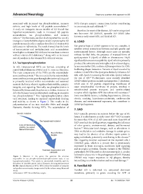
5.1. Tau hyperphosphorylation produced by astrocytes and microglia and, to a lesser degree,
In AD, intraneuronal NFTs are formed, consisting of by neurons in the CNS, mediates Aβ transport into the CNS,
paired helical filaments (PHFs) and 2.1 mm tau filaments. facilitating plaque formation. Variants of ApoE encoded by
The main components of the PHFs are the microtubule- codons 112 and 158 of the ApoE gene distinctly influence AD
associated tau protein. Tau is encoded by the microtubule- risk, with ApoEε4 increasing the risk while ApoEε2 reduces
54
58
associated tau protein gene through alternative splicing and the risk of AD. Furthermore, some recently identified
is primarily localized within microtubules and neuronal LOAD-related proteins include apolipoprotein J (clusterin),
axons in the brain, where it regulates their stability, synaptic which aids in Aβ peptide chaperoning; translocase of
integrity, and signaling. Normally, tau phosphorylation is outer mitochondrial membrane 40 protein, involved in
balanced by kinase and phosphatase activities. However, in mitochondrial protein transport; and sortilin-related
59
AD, this balance becomes disrupted, resulting in excessive receptor, which regulates APP interactions with secretases.
tau phosphorylation. This hyperphosphorylation alters Some modifiable factors, including hypertension, diabetes,
55
tau’s structure, leading to impaired microtubule binding obesity, smoking, hyperhomocysteinemia, cardiovascular
and stability, as shown in Figure 3. This results in the diseases, and environmental exposures, also contribute to
polymerization of tau into insoluble PHFs and straight LOAD pathogenesis.
filaments, thereby forming NFTs. The accumulation of
7. SAD
Genetic mutation SAD typically presents in patients before the age of 65,
hence, it is also known as early-onset AD. SAD accounts
60
for more than 95% of all AD cases and more than 60% of
Tau hyperphosphorylation
SAD cases lack the ApoE genotype, suggesting the influence
Loss of microtubule of other genetic, environmental, dietary, and hormonal
stability, tau paired factors. Molecular-level changes, such as alterations in
60
filament formation
Neurofibrillary DNA methylation and oxidative damage in certain genes,
tangles may lead to the absence of an effective repair system in
aging individuals, potentially contributing to the disease.
59
The triggering receptor expressed on the myeloid cells 2
(TREM2) gene, which is a protein that is prominently
expressed in brain microglia, modulates lipid signaling
and microglial activation. Genetic variants in the TREM2,
Neuronal degeneration and
cell death particularly R47H located on chromosome 6p21.1, are also
believed to play a role in the pathogenesis of SAD. Mutated
Figure 3. Mutation in the genes that generate the enzymes TREM2 may impair responses to Aβ plaques, thereby
responsible for tau phosphorylation/dephosphorylation leads to tau
hyperphosphorylation. This results in microtubule instability and the intensifying neuroinflammation and neuronal damage. 61
formation of insoluble paired helical filaments, forming intraneuronal
fibrillar deposits known as NFTs. NFTs reduce the number of synapses 8. Familial AD
and produce neurotoxicity. Image created by the authors using Microsoft
Word. Familial AD is inherited via autosomal dominant
Abbreviation: NFT: Neurofibrillary tangles. mutations, primarily in genes encoding for APP or
Volume 4 Issue 4 (2025) 22 doi: 10.36922/an.7087