
Page 11 - ARNM-3-1
P. 11
Advances in Radiotherapy
& Nuclear Medicine Diagnostics gude of biliary tract cancer
the attending physician and the patient’s specific condition. treatment effectiveness. In addition, USG aids in evaluating
These imaging methods have been extensively studied blood vessels in the digestive system and detecting fluid
for several decades, yet emerging techniques continue in the peritoneal cavity. It can also reveal changes in the
to provide new insights into the changes occurring in lymph nodes of the abdominal mesentery.
the patient’s body. Therefore, this review highlights the Ultrasound is one of the safest methods for diagnosing
most commonly used methods and discusses the types of digestive system issues, as its non-invasive nature allows
changes that can be observed through each technique.
for frequent repetition over short intervals without
2. Diagnostic methods for biliary tract posing any significant health risks to patients. The
cancer primary limitation, however, is the lack of a standardized
protocol for measuring and analyzing parameters during
2.1. Ultrasound an ultrasound examination. Transabdominal ultrasound
USG is a non-invasive, widely accessible diagnostic tool can confirm biliary dilation and exclude cholelithiasis,
32
commonly available in most medical facilities. It offers as well as detect masses in the liver. This technique is
33
a simple procedure without any harmful side effects, valuable in the early detection of biliary tract cancer. It
making it a highly favorable option for initial assessment. is particularly useful for the initial evaluation of tumor
Over recent years, intestinal USG has gained increasing masses within the bile ducts and gallbladder. On USG,
prominence, particularly in diagnosing inflammatory intrahepatic bile duct cancer can often appear as a mass
34
bowel diseases. Due to its numerous benefits, USG is lesion. eCCAs are typically echogenic, but they can still
34
now considered as valuable as more complex imaging be demonstrated and characterized using ultrasound.
techniques, such as CT. Unlike CT, USG does not expose Moreover, this examination allows for simple visualization
patients to ionizing radiation, further enhancing its of papillary tumors and nodular ductal carcinomas of the
appeal. biliary tract. 35,36 The extent of biliary tract involvement
can be assessed based on the location of the tumor and
During an ultrasound of the large intestine, clinicians the distribution of biliary obstruction. However, the
34
can detect not only changes within the intestinal wall specificity of ultrasound remains unknown. A 5-year
but also abnormalities beyond it. Key indicators, such population-based study conducted in Thailand showed
as variable echogenicity and increased Color-Doppler that USG can detect premalignant lesions and resectable
signals, may suggest excessive blood supply to tissue or biliary tract cancer at an early stage. For this reason, it is
decreased intestinal peristalsis. These features make USG recommended as the first screening tool for biliary tract
a useful tool for assessing disease activity, identifying cancer in patients aged ≥40 years in endemic areas.
37
complications, characterizing stenoses, and evaluating Technical improvements, such as the use of contrast-
enhanced ultrasound (CEUS) and the ability to perform
both transabdominal ultrasound and EUS, further expand
the potential of this imaging method in evaluating luminal
and extraluminal masses in the diagnosis of biliary tract
cancer. Rayubkul et al. showed that ultrasound screening
38
33
for gallbladder cancer improves early detection and may
reduce the need for expensive or invasive diagnostic
procedures. Similarly, Thinkhamrop et al. found that
39
ultrasound findings were strongly associated with CCA,
particularly in patients diagnosed with biliary dilation
and liver masses. In their study, of the 1,880 people who
underwent ultrasound and had a pathological diagnosis of
CCA, the overall detection rate was 35.74%. The detection
rate for those with liver masses was 54.85%, and for those
with dilated bile duct, it was 62.01%. Ultrasound is also
useful for guiding needle biopsy of intrahepatic lesions.
36
Advances in Doppler imaging and three-dimensional
ultrasound have further enhanced the application of
ultrasound in evaluating the biliary system. CEUS, which
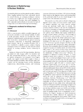
Figure 1. A map of cholangiocarcinoma imaging techniques combines traditional ultrasound with a contrast agent, has
Abbreviation: US: Ultrasound. emerged as a valuable tool. 40,41
Volume 3 Issue 1 (2025) 3 doi: 10.36922/arnm.4557