
Page 15 - ARNM-3-2
P. 15
Advances in Radiotherapy
& Nuclear Medicine Role of PET/CT in exploring tumor heterogeneity
A B C D
E F
G H
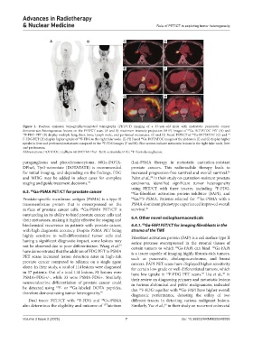
Figure 1. Positron emission tomography/computed tomography (PET/CT) imaging of a 33-year-old male with metastatic pancreatic cancer
demonstrates heterogeneous lesions on the PET/CT scan. (A and B) maximum intensity projection (MIP) images of Ga-DOTATOC-PET (A) and
68
68
18 F-FDG-PET (B) display multiple lung, liver, bone, lymph node, and peritoneal metastases. (C and D) Fused PET/CT of Ga-DOTATOC (C) and 18
F-FDG PET (D) display higher uptake of F-FDG in the right hilar node. (E–H) Fused Ga-DOTATOC images of the abdomen (E and G) display higher
68
18
uptake in liver and peritoneal metastases compared to the F-FDG images (F and H). Blue arrows indicate metastatic lesions in the right hilar node, liver
18
and peritoneum.
Abbreviations: DOTATOC: Gallium-68 (DOTA0-Phe1-Tyr3) octreotide; FDG: F-fluorodeoxyglucose.
18
paraganglioma and pheochromocytoma, 68Ga-DOTA- (Lu)-PSMA therapy in metastatic castration-resistant
DPhe1, Tyr3-octreotate (DOTATATE) is recommended prostate cancers. This radionuclide therapy leads to
for initial imaging, and depending on the findings, FDG increased progression-free survival and overall survival.
36
and MIBG may be added in select cases for complete Pabst et al., in their study on castration-resistant prostate
36
staging and guide treatment decisions. 33 carcinoma, identified significant tumor heterogeneity
using PET/CT with three tracers, including F-FDG,
18
6.3. Ga-PSMA PET/CT for prostate cancer 68 Ga-fibroblast activation protein inhibitor (FAPI), and
68
Prostate-specific membrane antigen (PSMA) is a type II 68 Ga/ F-PSMA. Patients selected for 177 Lu-PSMA with a
18
transmembrane protein that is overexpressed on the PSMA-dominant phenotype experienced improved overall
surface of prostate cancer cells. Ga-PSMA PET/CT is survival. 36
68
outstanding in its ability to bind prostate cancer cells and
their metastases, making it highly effective for staging and 6.4. Other novel radiopharmaceuticals
biochemical recurrence in patients with prostate cancer, 6.4.1. Ga-FAPI PET/CT for imaging fibroblasts in the
68
with high diagnostic accuracy. Despite PSMA PET being stroma of the TME
highly sensitive to well-differentiated tumor cells and Fibroblast activation protein (FAP) is a cell surface type II
having a significant diagnostic impact, some lesions may serine protease overexpressed in the stromal tissues of
not be observed due to poor differentiation. Wang et al. certain tumors to which Ga-FAPI can bind. Ga-FAPI
34
68
68
have demonstrated that the addition of FDG PET to PSMA is a tracer capable of imaging highly fibrosis-rich tumors,
PET scans increased lesion detection rates in high-risk such as pancreatic, cholangiocarcinoma, and breast
prostate cancer compared to reliance on a single agent cancers. FAPI PET scans have displayed higher sensitivity
alone. In their study, a total of 114 lesions were diagnosed for certain low-grade or well-differentiated tumors, which
in 37 patients. Out of a total 114 lesions, 81 lesions were have low uptake in F-FDG PET scans. Liu et al., in
18
38
37
PSMA+FDG+/-, while 33 were PSMA-FDG+. Similarly, their review on diagnosing primary and metastatic lesions
neuroendocrine differentiation of prostate cancer could in various abdominal and pelvic malignancies, indicated
be detected using F- or Ga-labeled DOTA peptides, that F-FDG together with Ga-FAPI have higher overall
68
18
68
18
therefore demonstrating tumor heterogeneity. diagnostic performance, denoting the utility of two
35
18
68
Dual tracer PET/CT with F-FDG and Ga-PSMA different tracers in detecting various malignant lesions.
also determines the eligibility and outcome of lutetium Similarly, Yue et al., in their study on recurrent colorectal
177
39
Volume 3 Issue 2 (2025) 7 doi: 10.36922/ARNM025040005