
Page 61 - BH-1-2
P. 61
Brain & Heart Enhancing peripartum cardiomyopathy awareness
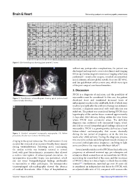
Figure 1. Electrocardiogram showing giant inverted T waves.
without any perioperative complications, the patient was
discharged and required to return for clinical and imaging
follow-up. Cardiac magnetic resonance imaging, which was
performed 1 month after surgery, revealed circumscribed
apical akinesia, efficient global systolic function (EF 60%),
and late gadolinium enhancement area, which were signs
of the post-surgical scar tissue formation.
3. Discussion
PPCM is a diagnosis of exclusion, and the possibility of
Figure 2. Transthoracic echocardiogram showing apical pedunculated myocarditis must be considered. In this case, the patient
endoventricular thrombus. developed fever and manifested increased levels of
inflammatory markers after childbirth, both of which were
A B resolved promptly after the antibiotic therapy was initiated;
therefore, a diagnosis associated with viral infection was
ruled out. The patient was considered having PPCM since
hypertrophy of the cardiac tissue occurred approximately
2 days after child delivery, falling within the time frame
where PPCM most commonly arises. The definitive
diagnosis was confirmed with myocardial biopsy, which
also did not reveal histopathological findings indicative of
myocarditis. PPCM is a growing entity, defined as a heart
failure-related cardiomyopathy that occurs classically
Figure 3. Cerebral computed tomography angiography. (A) Before during the last period of pregnancy or in the first few
procedure; (B) after stent retriever thrombectomy.
weeks after child delivery. During the diagnosis process,
it is crucial to exclude underlying heart muscle diseases or
clinging to the apical trabeculae. The small incision (3 cm) structural pathologies since pregnancy can bring to light
avoided the removal of an excessive healthy heart muscle some conditions that may resemble heart failure .
[2]
during ventriculostomy. Following aortic unclamping,
the cardiac activity was instantly restored to normal Several notable risk factors for PPCM are black ethnicity,
level, with good hemodynamic parameters that allowed pregnancy at advanced maternal age, pre-eclampsia and
a sudden interruption of extracorporeal circulation. An eclampsia. Black women have an increased risk for PPCM,
intraoperative myocardial biopsy was performed, which accounting for almost half of the cases, and the incidence
did not reveal histopathological findings attributable of PPCM is three to 4 times higher in black than in white
to myocarditis or other pathologies. The intraoperative women [8,9] . Furthermore, maternal age over 30 years old
transesophageal echocardiography showed the absence is considered an independent risk factor for PPCM, with
of residual thrombosis. After 9 days of hospitalization, an adjusted odds ratio of 1.8, compared with women who
Volume 1 Issue 2 (2023) 3 https://doi.org/10.36922/bh.1115