
Page 10 - BH-2-1
P. 10
Brain & Heart Hypochloremia in refractory heart failure
The osmotic gradient between the gut lumen and Kidneys are the key organs in HF-related congestion
mucosa is what causes fluid to be secreted into the intestinal and volume overload. They are the main organs affected
tract, and it is mainly caused by Cl and, to a lesser degree, by hypoperfusion and are the sites of primary counter-
HCO ions. Under normal circumstances, the kidneys regulatory responses. Moreover, the kidneys are a target
3
regulate the daily dietary intake of NaCl. The entire of prolonged diuretic therapy in patients with cardiac and
length of human kidney nephrons expresses Cl channels, renal disorders. However, the long-term use of diuretics
which take part in transepithelial transport, acidification results in decreased responsiveness and further renal
of intracellular vesicles, and cell volume regulation. The deterioration in the form of diuretic resistance. 26
macula densa in the juxtaglomerular part of the kidney
mainly uses Cl instead of Na to sense salt and volume status. 7. Diuretic resistance
When the volume is normal, there is less salt reabsorption There is no consensus in the literature among clinicians
in the proximal tubule, with more NaCl reaching the regarding the definition of diuretic resistance. However,
macula. This adequate NaCl sensed at the macula, in turn, it is agreed that the decreased diuretic and natriuretic
inhibits renin secretion and blocks RAAS. This effect is effects of loop diuretics worsen fluid overload. Diuretic
independent of Na; however, the effect does not occur resistance affects 25 – 30% of patients with HF and causes
when the macula is exposed to NA bicarbonate (NaHCO ) fluid retention despite higher doses of loop diuretics. 26,27
3
instead of NaCl. This neurohormonal mechanism suggests Many physiological alterations in CHF can cause changes
that Cl is mainly responsible for renin and volume status in drug pharmacokinetics, such as problems in drug
regulation, supporting the Cl theory. 23-25 absorption, distribution, metabolism, and excretion of
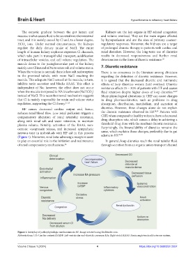
HF causes decreased cardiac output and, hence, diuretics. However, these changes alone do not explain
reduces renal blood flow. Low renal perfusion triggers a the diuretic resistance observed in HF. 28,29 Patients with
compensatory alteration of renal arteriolar resistance, CHF, when compared to healthy subjects, have a decreased
along with renal salt and water retention, to maintain drug absorption rate, which causes a delay in achieving a
plasma volume. Notably, activation of the RAAS, non- threshold drug dose with the resultant diuretic resistance.
osmotic vasopressin release, and increased sympathetic Surprisingly, the bioavailability of diuretics remains the
nervous tone in individuals with HF aid in this process same, which explains these changes, preferably due to gut
29,30
(Figure 1). Moreover, renal beta-adrenergic receptors seem edema in HF.
to play an essential role in the initiation and maintenance In general, loop diuretics reach the renal tubular fluid
of renal compensatory mechanisms. 26 through secretion from an organic anion transport channel
Figure 1. Interplay of pathophysiologic mechanisms in HF. Image created using BioRender.com.
Abbreviations: CO: Cardiac output; LVEDP: Left ventricular end-diastolic pressure; RA: Right atrial; RAAS: Renin angiotensin aldosterone system.
Volume 2 Issue 1 (2024) 4 https://doi.org/10.36922/bh.2257