
Page 24 - BH-3-3
P. 24
Brain & Heart Modern imaging and management of bicuspid valves
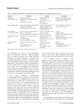
Table 1. Strengths and limitations of multi‑modality cardiac imaging for evaluating bicuspid aortic valves
Imaging Indications Strengths Limitations
Transthoracic • Initial evaluation of BAV • Widely available and • Limited acoustic window in some
echocardiography (TTE) • Assessing valve morphology, stenosis, or cost-effective patients (obesity, chest deformities)
regurgitation • High temporal resolution • Suboptimal aortic imaging
• Aortic root measurements • Non‑invasive
• Real‑time imaging
Stress echocardiography • Assessing functional capacity in BAV with • Evaluates dynamic changes in • May not be suitable for all patients
moderate-to-severe stenosis gradient or regurgitation (e.g., severe BAV stenosis)
• Identifying exercise‑induced changes • Simultaneous functional and
structural assessment
Transesophageal • Detailed evaluation of valve morphology • Superior image quality for valve • ‑invasive
echocardiography (TEE) • When TTE is inconclusive and proximal aorta • Requires sedation
• Assessing endocarditis or aortic dissection • High spatial resolution • Limited in evaluating the distal aorta
Computed tomography • Evaluation of the ascending aorta and • Excellent spatial resolution • Radiation exposure
(CT) coarctation • 3D reconstruction • Requires contrast (limitation in renal
• Pre‑surgical planning • Superior for aortic geometry and dysfunction or allergies)
• Calcium quantification in stenosis measurements
Magnetic resonance • Assessment of ascending aorta size and • No radiation exposure • Expensive
imaging function • Superior for aortic flow • Limited availability
• Valve‑related flow dynamics quantification • Contraindications in patients with
• Longitudinal follow‑up (e.g., regurgitant fraction) certain implants or devices
• 3D imaging
Abbreviations: 3D: Three-dimensional; BAV: Bicuspid aortic valve.
valve calcification, the presence of aneurysmal disease, due to its semi-invasive nature. It should be reserved for
and the interpreter’s experience. Three-dimensional selected cases, such as those undergoing AVR, where
TTE can improve detection rates. When significant precise preoperative morphological assessment is crucial.
calcification or stenosis complicates a bicuspid valve, TTE is typically sufficient to diagnose a BAV
accurately determining the number of leaflets becomes (Figure 1) and evaluate associated AS and AR valve
more challenging. In cases of severe AS requiring valve dysfunction. Patients with a BAV should undergo Doppler
replacement, TTE becomes less reliable in determining the echocardiography to assess for AS, measure the diameters
number of valve leaflets. In a study of 100 patients (mean of the aortic root and thoracic aortic aneurysm to check for
age 70 years) with AS undergoing valve replacement, dilation, and evaluate the presence of aortic coarctation. 3,39,40
echocardiograms were interpretable in 86 cases. Of these, Echocardiography plays a crucial role in assessing the
35
44 patients had congenitally malformed valves, and 42 had severity of AS, utilizing Doppler techniques to measure the
tricuspid valves, with echocardiographic findings matching peak velocity and mean gradient across the stenotic aortic
valve structure in only 66% of cases (57 out of 86). valve. These measurements, along with the calculation of
Given these limitations, transesophageal the aortic valve area (AVA) using the continuity equation,
echocardiography (TEE) can provide additional value in help determine the severity of AS. 41,42 . Echocardiography
evaluating aortic valve morphology. Several studies have also provides insight into left ventricular function, such as
demonstrated the superiority of two-dimensional TEE ejection fraction and the presence of hypertrophy, which
over two-dimensional TTE in accurately diagnosing valve may result from the increased afterload caused by AS. The
phenotypes. For instance, a study by Chu et al. found strengths of echocardiography include its non-invasive
37
that the sensitivity and specificity of TEE for diagnosing nature, widespread availability, and ability to assess both
unicuspid aortic valves were 75% and 86%, respectively, the aortic valve and left ventricle in real-time. However,
compared to 27% and 50% for TTE. This underscores TEE’s limitations include the potential for underestimating
greater reliability in distinguishing the unicuspid aortic AS severity in low-flow states, challenges in accurately
valve from BAV. In addition, Yousry et al. reported that measuring AVA with eccentric jets, and difficulties in
37
38
TEE had a higher interobserver agreement, sensitivity, obtaining optimal Doppler signals due to poor acoustic
42,43
and specificity than TTE for evaluating valve phenotype, windows.
reinforcing its utility in challenging diagnostic scenarios. Echocardiography is a vital tool for assessing the severity
38
Nevertheless, TEE is not recommended for all patients of AR, allowing for both qualitative and quantitative
Volume 3 Issue 3 (2025) 5 doi: 10.36922/BH025050008