
Page 21 - BH-3-3
P. 21
Brain & Heart Modern imaging and management of bicuspid valves
ago. Early recognition of BAV is crucial, as physicians
1
have become increasingly aware of its lifelong impact on
cardiovascular health. BAV is now thought to be more
2
of a syndrome rather than an isolated valvular pathology.
The presence of a bicuspid valve not only increases the risk
of valvular complications such as stenosis, regurgitation,
and infection but is also associated with aortopathies,
including aneurysms and acute aortic syndromes. This
has led to heightened interest among investigators to try
to comprehensively understand the pathology of BAV and
its diverse implications, and regularly updated guidelines
in the evaluation and treatment of these patients. This
3
review aims to provide a contemporary overview of clinical
perspectives, multi-modality imaging, and management
strategies for BAVs.
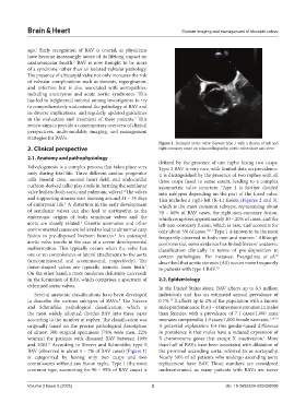
Figure 1. Bicuspid aortic valve (Sievers type 1 with a fusion of left and
2. Clinical perspective right coronary cusp) on echocardiography, aortic valve short axis view
2.1. Anatomy and pathophysiology
defined by the presence of one raphe fusing two cusps.
Valvulogenesis is a complex process that takes place very Type 2 BAV is very rare, with limited data on prevalence.
early during fetal life. Three different cardiac progenitor It is distinguished by the presence of two raphes with all
cells (neural crest, second heart field, and endocardial three cusps fused to some extent, leading to a complex
cushion-derived cells) play a role in forming the semilunar asymmetric valve structure. Type 1 is further divided
valve leaflets (both aortic and pulmonic valves). The valves into subtypes depending on the part of the fused valve.
4
and supporting sinuses start forming around 31 – 35 days This includes a right-left (R–L) fusion (Figures 2 and 3),
of embryonic life. A distortion in the early development which is the most common subtype, representing about
5
of semilunar valves can also lead to aortopathy, as the 70 – 80% of BAV cases, the right-non-coronary fusion,
embryonic origins of both semilunar valves and the which comprises approximately 10 – 20% of cases, and the
aorta are closely related. Genetic anomalies and other left-non-coronary fusion, which is rare, and accounts for
4
environmental causes are believed to lead to abnormal cusp only about 5% of cases. 10,11 Type 1 is known to be the most
fusion in pre-disposed liveborn humans. An unicuspid frequently observed in both men and women. Although
6
1
aortic valve results in the case of a severe developmental controversial, some evidence has linked Sievers’ anatomic
malformation. This typically occurs when the valve has classification clinically in terms of pre-disposition to
one or no commissures or lateral attachments to the aorta certain pathologies. For instance, Evangelista et al.
12
(unicommissural and acommissural, respectively). The described that aortic stenosis (AS) occurs more frequently
dome-shaped valves are typically stenotic from birth. in patients with type 1 BAV. 12
7
On the other hand, a more moderate deformity can result
in the formation of BAV, which comprises a spectrum of 2.2. Epidemiology
deformed aortic valves. In the United States alone, BAV affects up to 6.5 million
Several anatomic classifications have been developed individuals and has an estimated annual prevalence of
to describe the various subtypes of BAVs. The Sievers 0.7%. It affects up to 2% of the population with a known
13
8
and Schmidtke pathological classification, which is male predominance. It is 3 – 4 times more common in males
the most widely adopted, divides BAV into three types than females, with a prevalence of 7.1 cases/1,000 male
according to the number of raphes. The classification was neonates compared to 1.9 cases/1,000 female neonates. 1,13,14
originally based on the precise pathological description A potential explanation for this gender-based difference
of almost 300 surgical specimens (78% were men, 22% in prevalence is that males have a reduced expression of
women) for patients with diseased BAV between 1999 X chromosome genes that escape X inactivation. More
1
and 2003. According to Sievers and Schmidtke, type 0 than half of BAVs have been associated with dilatation of
9
BAV (observed in about 5 – 7% of BAV cases) (Figure 1) the proximal ascending aorta, referred to as aortopathy.
is categorized by having only two cusps and two Nearly 50% of all patients who undergo ascending aorta
commissures without any fusion raphe. Type 1 (the most replacement have BAV. Those numbers are considered
common type, accounting for 90 – 95% of BAV cases) is underestimated, as many patients with BAVs are never
Volume 3 Issue 3 (2025) 2 doi: 10.36922/BH025050008