
Page 157 - EJMO-9-3
P. 157
Eurasian Journal of
Medicine and Oncology Pfannenstiel incision in endometrial cancer
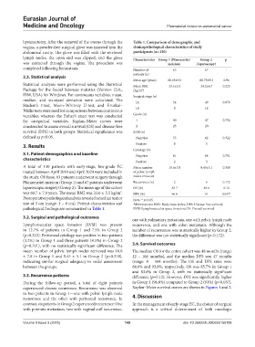
hysterectomy. After the removal of the uterus through the Table 1. Comparison of demographic and
vagina, a powder-free surgical glove was inserted into the clinicopathological characteristics of study
abdominal cavity. The glove was filled with the retrieved participants (n=130)
lymph nodes, the open end was clipped, and the glove Characteristics Group 1 (Pfannenstiel Group 2 p
was extracted through the vagina. The procedure was incision) (laparoscopy)
completed following hemostasis. Number of 63 67 -
patients (n)
2.3. Statistical analysis
Mean age (years) 60.65±9.5 60.75±9.2 0.96
Statistical analyses were performed using the Statistical Mean BMI 33.1±5.5 34.2±4.7 0.225
Package for the Social Sciences statistics (Version 22.0., (kg/m )
2
IBM, USA) for Windows. For continuous variables, mean, Surgical stage (n)
median, and standard deviation were calculated. The IA 54 49 0.078
Student’s t-test, Mann–Whitney U-test, and Kruskal–
Wallis tests were used for comparisons between continuous IB 9 18
variables, whereas the Fisher’s exact test was conducted Grade (n)
for categorical variables. Kaplan–Meier curves were 1 40 47 0.702
constructed to assess overall survival (OS) and disease-free 2 23 20
survival (DFS) in both groups. Statistical significance was LVSI (n)
defined as p<0.05. Negative 55 62 0.322
3. Results Positive 8 5
Cytology (n)
3.1. Patient demographics and baseline Negative 61 64 0.701
characteristics
Positive 2 3
A total of 130 patients with early-stage, low-grade EC Mean number 10.6±7.8 9.45±5.1 0.318
treated between April 2010 and April 2024 were included in of pelvic lymph
the study. Of these, 63 patients underwent surgery through nodes removed
Pfannenstiel incision (Group 1) and 67 patients underwent Recurrence (n) 2 6 0.172
laparoscopic surgery (Group 2). The mean age of the cohort OS (%) 85.7 83.6 0.12
was 60.7 ± 7.9 years. The mean BMI was 33.6 ± 5.5 kg/m . DFS (%) 96.8 91 0.037*
2
Postoperative pathological analysis revealed a median tumor Note: * p<0.05.
size of 3 cm (range: 1 – 8 cm). Patient characteristics and Abbreviations: BMI: Body mass index; DFS: Disease-free survival;
pathological findings are summarized in Table 1. LVSI: Lymphovascular space invasion; OS: Overall survival.
3.2. Surgical and pathological outcomes
one with pulmonary metastasis, one with pelvic lymph node
Lymphovascular space invasion (LVSI) was present recurrence, and one with colon metastasis. Although the
in 12.7% of patients in Group 1 and 7.5% in Group 2 number of recurrences was numerically higher in Group 2,
(p=0.322). Peritoneal cytology was positive in two patients the difference was not statistically significant (p=0.172).
(3.2%) in Group 1 and three patients (4.5%) in Group 2
(p=0.701), with no statistically significant difference. The 3.4. Survival outcomes
mean number of pelvic lymph nodes retrieved was 10.6 The median OS for the entire cohort was 48 months (range:
± 7.8 in Group 1 and 9.45 ± 5.1 in Group 2 (p=0.318), 12 – 168 months), and the median DFS was 47 months
indicating similar surgical adequacy in nodal assessment (range: 8 – 168 months). The OS and DFS rates were
between the groups. 84.6% and 93.8%, respectively. OS was 85.7% in Group 1
and 83.6% in Group 2, with no statistically significant
3.3. Recurrence patterns difference (p=0.12). However, DFS was significantly higher
During the follow-up period, a total of eight patients in Group 1 (96.8%) compared to Group 2 (91%) (p=0.037).
experienced disease recurrence. Recurrence was observed Kaplan–Meier survival curves are shown in Figures 1 and 2.
in two patients in Group 1—one with pelvic lymph node
recurrence and the other with peritoneal recurrence. In 4. Discussion
contrast, six patients in Group 2 experienced recurrence: One In the management of early-stage EC, the choice of surgical
with port-site metastasis, two with vaginal cuff recurrence, approach is a critical determinant of both oncologic
Volume 9 Issue 3 (2025) 149 doi: 10.36922/EJMO025150106