
Page 96 - GPD-2-2
P. 96
Gene & Protein in Disease Immunoblotting large and small proteins
A B
C D
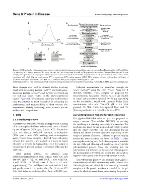
Figure 1. (A) Density plot of human proteome based on Uniprot data. Protein mass in logarithmic scale is represented and highlighted for SVIP and MGA.
(B) Summary workflow for a western blot using the SWP and their adaptations for small and large proteins. Examples of western blots against (C) SVIP
and (D) MGA performed modifying the standard protocol as shown in B. SVIP western blot was performed on cell lysates of BB30-HNC cell line either
transfected with pLVX-ZSgreen empty vector (EV) or containing SVIP coding sequence (CDS). MGA western blot was performed on cell lysates of
LouNH91 as negative control (NC), and HEK 293T transfected with pLVX-ZSgreen containing MGA CDS.
Abbreviations: NFSM Non-fat skim milk; SWP: Standard working procedure; SVIP: Small VCP-interacting protein; MGA: MAX-gene-associated protein.
These changes were used in different studies involving Collected supernatants are quantified through the
™
small VCP-interacting protein (SVIP) and MAX-gene- Lowry method using the DC Protein Assay Kit II
[4]
[7]
associated protein (MGA) [5,6] , and they led to overcoming (BioRad, 5000112). Then, samples are prepared for
the technical issues related to the above-mentioned electrophoresis. Quantified protein extracts are diluted
targets (Figure 1B). We anticipate that these modifications to equal concentrations (around 10–30 µg depending
have the potential to assist researchers in enhancing the on the availability), mixed with Laemmli buffer (final
performance and reproducibility of their western blot concentration: 62.5 mM Tris-HCl, pH = 6.8, 25%
experiments, thereby facilitating more accurate protein glycerol, 2% SDS, 0.01% bromophenol blue, and 5%
analysis and interpretation. β-mercaptoethanol) and boiled for 5 min at 95°C.
2. SWP 2.2. Electrophoresis-mediated protein separation
2.1. Sample preparation Tris glycine-SDS-Polyacrylamide gels are prepared in
empty cassettes (Thermofisher, NC2015) by pouring
Adherent cells are collected using a scrapper after washing the resolving and stacking mixes into the cassettes. The
cells in phosphate-buffered saline (PBS) twice, followed resolving mix must be first prepared and rapidly poured
by centrifugation (2000 rpm, 2 min, 4°C). Suspension into the empty cassettes. This mix determines the gel
cells are directly collected through centrifugation density and allows a proper separation depending on the
(2000 rpm, 2 min, 4°C), washing, and centrifugation weight of protein of interest. Different recipes of varying
again. Frozen tissue samples – derived from orthotopic acrylamide concentrations are listed in Table 1. After
tumors, for instance – are mashed in a pestle in liquid pouring the resolving mix, 2 mL of isopropanol is added to
nitrogen to prevent its degradation. Once the sample is the top of the gel. This step will accelerate the acrylamide
homogenized, protein extract is obtained following the polymerization process. Once the resolving mix has
next steps. polymerized and followed by the removal of isopropanol,
Total protein extracts are obtained using the stacking mix is prepared and loaded into the cassettes,
radioimmunoprecipitation assay (RIPA) buffer (20 mM together with a comb with the desired number of wells.
Tris-HCl [pH = 7.5], 150 mM NaCl, 1 mM Na EDTA, The polymerized gel is set in a mini gel tank (A25977,
2
1 mM EGTA, 1% NP-40) (100 µL for 2 × 10 cells, Thermofisher) and filled with running buffer (25 mM Tris,
6
approximately). Then, cell lysates are sonicated for 5 min 200 mM glycine, and pH = 8.5). After removing the comb,
and gently mixed in the rotator wheel for 15 min at 4°C. samples and the protein markers are loaded in the gel,
After that, tubes are centrifuged for 15 min at 13,000 rpm. where the electrophoresis takes place, forcing the proteins
Volume 2 Issue 2 (2023) 2 https://doi.org/10.36922/gpd.0547