
Page 82 - GTM-1-1
P. 82
Global Translational Medicine Quantification of atherosclerosis
2. En face analysis of atherosclerotic lesions A B
C
En face method is used routinely for the entire length of the
aorta from the ascending to the abdominal aortic regions
above the iliac bifurcation (Figure 1A). After dissection
from the mouse, the aorta is fixed in either 10% neutrally
buffered formalin or 4% paraformaldehyde for at least 24 h
to preserve the tissue. Adventitial tissues are then removed
carefully and the aorta is cut open longitudinally through
the inner curvature and down the anterior aspect. The
aortic arch has three major branches: Innominate artery, D
left common carotid artery, and left subclavian artery
(Figure 1A). These three branches have been used as
landmarks to cut open and flat the aorta through the outer
curvature. In the published literature, there have been two
major modes to cut open the three aortic branches. The
first mode is to cut open and retain the innominate and
left carotid arteries but cutoff the left subclavian branch,
and use its orifice to open the aorta (Figure 1B and C). The E F
second mode is to cut open and retain all three branches,
and then make an additional cut of the outer curvature
in the descending thoracic aorta (Figure 1D). Although
many published articles use the second mode shown in
Figure 1D, we recommend using the first method as shown
in Figure 1B and C unless researchers want to focus on en
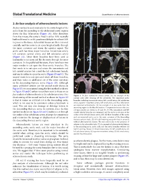
face analysis of atherosclerosis in the subclavian artery. One Figure 1. En face method for mouse aortas. (A) An example of the
shortcoming of the second method as shown in Figure 1D normal aorta after cleaning off the adventitia: The important aortic
is that it makes an artificial cut in the descending aorta, branches including innominate artery, left carotid artery, left subclavian
which is not easy to be consistent unless a landmark is artery, superior mesenteric artery, left renal artery, and iliac bifurcation
used. This cut may also damage or dislodge lesions in are remained as landmarks. (B) An example of en face aorta from the
the descending thoracic aorta. In contrast, since the first ascending region to the iliac bifurcation. Comparisons between the two
cut-open modes are shown in (C and D). (C) The innominate and left
method as shown in Figure 1B and C makes a cut through carotid arterial branches are remained, and the aorta is cut open at the
the orifice of the subclavian artery, it keeps the consistency orifice of the left subclavian artery. (D) All three branches of the aortic
and minimizes the damage or displacement of lesions in arch are remained and a cut to the outer curvature of the descending
the descending thoracic aortic region. thoracic aorta is made. (E) Oil red O staining was performed in an aorta
without atherosclerotic lesions. The red color is due to the presence of
Atherosclerotic lesions are most abundant in the adipose in the adventitial side. (F) An example of atherosclerotic lesions
ascending aorta, aortic arch, and the major branches of in the ascending aorta, aortic arch, and the aortic branches without Oil
the aortic arch. Therefore, it is important to be extremely Red O staining. Notes: (1) Innominate artery, (2) left carotid artery, (3)
left subclavian artery, (4) superior mesenteric artery, (5) left renal artery,
careful when cutting open the aorta, which should be and (6) iliac bifurcation.
performed under a dissecting microscope. The aorta
should be immersed in either saline or phosphate-buffered
saline to prevent the vessel from drying out. Fine tipped as atherosclerotic plaques. Furthermore, some lesions may
(tip diameter ~ 0.05 mm) Vannas spring scissors should be fragile and can be displaced during the staining process.
be used for opening the aortic branches due to their small This is particularly the case for lesions in mice that have
size. We suggest that 1 time users practice using normal undergone bone marrow transplantation. Considering
st
aortas and master the technique before performing an these issues, there is no tangible benefit to performing
atherosclerosis study. staining at least for large and mature plaques (Figure 1F),
and in fact, there may be some detriments.
Oil red O staining has been frequently used for en
face analysis of atherosclerosis. Although the red color Some software packages provide functions to
enhances the visualization of lesions, it is worth noting recognize and automatically measure atherosclerotic
that this method also stains neutral lipid of adipose in the lesion areas. However, even with images captured using
adventitia (Figure 1E), which may be mistakenly evaluated a high-resolution microscope, it cannot completely avoid
Volume 1 Issue 1 (2022) 2 https://doi.org/10.36922/gtm.v1i1.76