
Page 68 - GTM-2-2
P. 68
Global Translational Medicine Modified cardiac catheterization
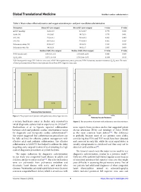
Table 3. Mean values of hemodynamics and oxygen saturation pre‑ and post‑vasodilator administration
Parameters Mean±SD (pre‑oxygen) Mean±SD (post‑oxygen) t‑value P‑value
mPAP (mmHg) 36.8±14.9 41.5±14.7 0.778 0.445
Aorta (%) 93.2±4.5 98.7±2.3 3.770 0.001
SVC (%) 73.2±14.1 78.3±15.2 0.852 0.403
IVC (%) 68.7±16.4 77.3±26.3 0.961 0.347
Pulmonary artery (%) 82.8±14.7 93.0±3.0 2.355 0.029
Pulmonary vein (%) 96.3±2.8 99.0±1.7 2.855 0.009
Median (IQR) (Pre‑oxygen) Median (IQR) (Post‑oxygen) U‑value P‑value
PVR (woods unit) 0.92 (0.3–1.2) 1.35 (0.68–4.25) 8.000 0.569
Q /Q ratio 3.27 (1.1–9.0) 2.75 (2.46–3.37) 11.500 0.919
P S
IQR: Interquartile range; IVC: Inferior vena cava; mPAP: Mean pulmonary artery pressure; PVR: Pulmonary vascular resistance; Q /Q ratio: The ratio
S
P
of total pulmonary blood flow to total systemic blood flow; SVC: Superior vena cava
Figure 1. The proportion of patients with pulmonary artery hypertension.
a tertiary healthcare center in Ibadan only reported its Figure 2. The proportion of patients with pulmonary vascular resistance.
initial diagnostic catheterization experience in children .
[3]
Rwebembera et al. in Uganda reported collaboration some reports from previous studies that suggested patent
between adult and pediatric cardiac interventional teams ductus arteriosus (PDA) and tetralogy of Fallot (TOF)
for diagnostic and therapeutic cardiac catheterization . as the most common heart defects [2-4] . The difference
[1]
The report suggested that catheterization procedures can is probably because most of our patients were adults,
be fully utilized for effective patient management with considering that PDA and TOF would have been detected
effective adult and pediatric collaboration. The effective and corrected in early life, while the atrial septal defect is
collaboration in LASUTH has helped to address the delay usually asymptomatic in childhood and thus may not be
in getting early surgical treatment by eliminating the high detected until adulthood .
[14]
costs of diagnostic procedures at private facilities. The femoral vessels were the major access used for our
The major indication for diagnostic catheterization diagnostic catheterization, similar to a previous study .
[2]
in our study was congenital heart disease in adults and Only one of the patients had internal jugular access because
children, similar to earlier studies [2-4] . The other indications of associated persistent left superior vena cava that might
were cor pulmonale from pulmonary embolism and pose difficulty in accessing the pulmonary artery. Two of
rheumatic heart disease with aortic and mitral valve our patients had additional diagnoses of other congenital
diseases. Ventricular and atrial septal defects were the most heart abnormalities missed on echocardiography,
common congenital heart defect, which is at variance with which included persistent left superior vena cava and
Volume 2 Issue 2 (2023) 4 https://doi.org/10.36922/gtm.249