
Page 75 - GTM-2-2
P. 75
Global Translational Medicine Elastolysis of porcine aortic adventitia
found [40,41] that human neutrophil elastase (HNE) degraded was subjected to 1-h elastolysis, either without or with
collagen Type I from pig tendon or rabbit skin, collagen subsequent irradiation. The photochemical crosslinking
Type II from pig cartilage, and collagen Type IV from the of collagen is likely responsible for the mechanical
kidney basement membrane. It was suggested that the augmentation of adventitia, whether degraded in the
elastolytic attacks occurred either at terminal peptides or short- or long-term. However, in the latter case, the high
in the helical region. Since then, other investigators have level of degradation has allowed only a limited mechanical
reported elastolysis with HNE of collagens types III [42,43] , recovery. We can only conclude that such extensive
IX, X, and XI , IV , and I . enzymolytic damage can be explained by the degradation
[44]
[45]
[46]
of collagen by elastase, as no collagenase was present in the
3.2. Effect of irradiation system.
Figure 1 shows that the exposure to UV-A radiation of
the degraded adventitial specimens led to an increase 3.4. Effects on adventitial/medial tissue
in both stiffness and strength of the tissue, likely due Figure 3A presents a section through the native bovine
to the additional covalent crosslinks generated in the aortic wall before degradation and/or irradiation.
photochemical process. The enhanced mechanical Residual medial tissue can be seen in all samples. The
properties clearly show that UV-irradiation is beneficial devastating effect of the long-term elastolysis (48 h) is
to an already degenerated wall at any stage before the clearly illustrated in Figure 3B: elastin’s presence (black
aneurysmal rupture; the earlier, the better. Presumably, stain) was reduced massively in the media and completely
the collagen crosslinking strengthens and stiffens the digested in the adventitia, while the collagen network
adventitia sufficiently to delay a potential rupture. We (red stain) became disorganized and depleted. It is
reached the same conclusion in a previous in vitro study evident that elastase can digest collagen. The short-term
based on experimental collagenolysis . elastolysis (1 h) had a surprisingly drastic effect on the
[22]
amount of elastin, with only little left in the adventitia
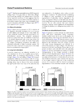
3.3. Long-term elastolysis (Figure 3C, arrows) and significantly depleted in media.
In another experiment, we subjected adventitia to in Disorganization of the collagen network is already
vitro elastolysis for 48 h. This lengthier process had a obvious in both degraded tissues. Figure 3D shows the
dramatic effect on adventitial biomechanics, as both effect of irradiation with UV-A rays of a specimen that
strength (Figure 2A) and stiffness (Figure 2B) dropped was subjected beforehand to 1-h elastolysis. It can be seen
considerably. Following the irradiation with UV-A rays, that irradiation induced a more compact fine structure
these characteristics recovered to some extent, eventually of the collagen network, likely due to the crosslinking
reaching values close to those of the native adventitia process. It is consistent with the observed mechanical
but significantly lower than those of the adventitia that augmentation of the adventitial samples and confirms
A B
Figure 2. The effect of elastolysis duration on the mechanical properties of isolated tunica adventitia of the porcine abdominal aorta, before and after
irradiation with ultraviolet-A rays (365 nm, 45 mW/cm , 10 min), measured for n = 30 in each set of samples. The bar graphs present a comparative
2
evaluation of (A) ultimate tensile stress (strength) and (B) Young’s modulus (stiffness) between specimens digested for either 1 h or 48 h in elastase, as
measured uniaxially in an Instron Model #5943 mechanical tester. High statistical differences can be observed between the effects of the two different
durations of the experimental elastolysis.
Volume 2 Issue 2 (2023) 5 https://doi.org/10.36922/gtm.0897