
Page 94 - GTM-3-1
P. 94
Global Translational Medicine Late abscess complication endometrial cancer
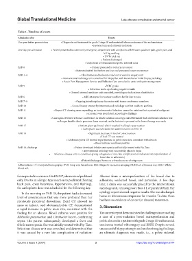
Table 1. Timeline of events
Admission day Events
One year before presentation • Diagnosis and treatment for grade 2 stage IB endometrioid adenocarcinoma of the endometrium
• Hysterectomy and adjuvant radiation
One day pre-admission • Patient presented to community emergency department with complaints of left lower quadrant pain, groin pain, and
left leg swelling
• DVT ruled out
• Patient discharged
• Outpatient CT demonstrated pelvic sidewall mass
PAD 0 • Patient presented to tertiary care center
• Patient admitted for further work up and presumed cancer recurrence
PAD 1–4 • Claudication and ischemia ruled out at vascular surgery unit
• Interventional radiology unit consulted for biopsy but with inconclusive initial biopsy pathology
• Acute Pain Management Service and Palliative Care consulted to assist with pain management
PAD 5 • WBC peaks
• Infectious work‑up showing negative results
• General internal medicine unit consulted, providing no indications of antibiotics
PAD 6 • MRI attempted but patient unable to lie flat due to pain
PAD 7–9 • Ongoing interdisciplinary discussion with tumor conference members
PAD 10 • Second biopsy attempt by interventional radiology unit but unable to perform
PAD 11 • Repeat CT showing mass expanding; an involvement of infection cannot be ruled out but a potential malignant
recurrence was speculated, according to findings
PAD 15 • Case again reviewed at tumor conference, in which radiation oncology unit determined that additional radiation was
no longer feasible due to previous doses received, and a decision to proceed with chemotherapy was made
PAD 17 • Patient given paclitaxel, which resulted in allergic‑type reaction
• Carboplatin was scheduled for administration on PAD 18
PAD 18 • Significant decrease in level of consciousness
• Head CT was normal
• Abdomen/pelvis CT showed rapid increase in pelvic mass size, consistent with abscess
• Blood cultures result was positive
PAD 18–discharge • Patient developed febrile neutropenia and initially treated with Pip‑Tazo
• Interventional radiology unit successfully placed a drain
• Infectious disease unit re‑consulted, giving a diagnosis of rare, late complication of radiation (microperforation of
bowel due to adhesions)
• Patient discharged home on a 4‑week course of ertapenem
Abbreviations: CT: Computed tomography; DVT: Deep vein thrombosis; MRI: Magnetic resonance imaging; PAD: Post-admission day; WBC: White
blood cell.
for suspected recurrence. On PAD 17, she received paclitaxel Abscess from a microperforation of the bowel due to
only. Due to an allergic-type reaction to paclitaxel (fleeting adhesions, weakened bowel, and peristalsis. A few days
back pain, chest heaviness, hypertension, and flushing), later, a drain was successfully placed by the interventional
the carboplatin dose was scheduled for the following day. radiology unit, releasing more than 1 L of purulent fluid. Her
In the morning on PAD 18, the patient had a decreased cytology report showed negative results. She was discharged
level of consciousness that was more profound than her home on intravenous ertapenem for 4 weeks. To date, there
previously perceived drowsiness. Head CT showed no has been no evidence of cancer (or abscess) recurrence.
mass or infarct, and abdomen/pelvis CT demonstrated 3. Discussion
a rapid increase in pelvic mass size, consistent with the
finding for an abscess. Blood cultures were positive for The current report demonstrates the challenges in unraveling
Klebsiella pneumoniae and Citrobacter koseri, confirming a case of a post-radiation bowel microperforation and
sepsis. The patient subsequently developed a fever and pelvic abscess in a patient with grade 2 stage 1B endometrial
febrile neutropenia. She was initially treated with Pip-Tazo. carcinoma treated with surgery and EBRT. Despite several
Infectious disease unit was consulted and determined that unsuccessful biopsy attempts and unclear imaging findings,
it was caused by a rare late complication of radiation: an ultimate diagnosis was made, i.e., a pelvic sidewall
Volume 3 Issue 1 (2024) 3 https://doi.org/10.36922/gtm.2114