
Page 71 - GTM-3-2
P. 71
Global Translational Medicine Genes and blood cells in Ph-negative MPNs
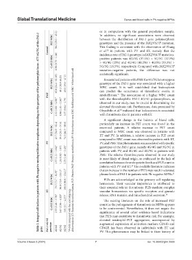
Table 2. Integral cell indices in patients with Ph-negative myeloproliferative neoplasms depending on the genotype of the polymorphic locus −675 4G/5G of the PAI-1
In addition, no significant associations were observed
PLT/WBC 38.2 (29.3 – 45.2) or in comparison with the general population sample.
between the distribution of PAI-1 gene polymorphism
genotypes and the presence of the JAK2V617F mutation.
Control This finding is consistent with the observation of Zhang
et al. in patients with PV and ET, namely that the
20
incidence rate of PAI-1 genotype in JAK2V617F mutation-
C×PLT/RBC 0.96 (0.79 – 1.16) positive patients was 4G/5G (37.5%) = 5G/5G (37.5%)
> 4G/4G (25%) and 4G/4G (48.2%) > 4G/5G (33.3%) >
5G/5G (18.5%), respectively. Compared with JAK2V617F
mutation-negative patients, the difference was not
statistically significant.
In examined patients with PMF, the 4G/5G heterozygous
PLT/WBC 62.8 (24.4 – 81.65) 40.2 (30.0 46.25) 45.0 (19.6 – 47.2) genotype of the PAI-1 gene was associated with a higher
WBC count. It is well established that leukocytosis
PMF can predict the occurrence of thrombotic events in
myelofibrosis. The association of a higher WBC count
27
with the thrombophilic PAI-1 4G/5G polymorphism, as
C×PLT/RBC 2.52 (1.06 – 4.58)* 1.84 (1.45 – 2.36)* 1.37 (0.64 – 1.79) observed in our study, may be crucial in determining the
elevated thrombosis risk. Furthermore, data presented by
Ohyashiki et al. indicated that leukocytosis is associated
28
with thrombosis also in patients with ET.
A significant change in the balance of blood cells,
particularly an increase in PLT count, was found in the
PLT/WBC 43.7 (34.0 – 62.4) 39.4 (33.3 – 57.6) 74.5 (44.3 – 89.6) examined patients. A relative increase in PLT count
compared to WBC count was observed in patients with
ET and PV. In addition, a relative increase in PLT count
PV compared to RBC count was observed in patients with ET,
PV, and PMF. This phenomenon was associated with specific
genotypes of the PAI-1 gene, namely 4G/4G and 5G/5G in
C×PLT/RBC 1.57 (1.11 – 1.64)* 1.24 (0.82 – 1.67) 2.23 (1.48 – 2.43)* Abbreviations: ET: Essential thrombocythemia; PLT: Platelet; PMF: Primary myelofibrosis; PV: Polycythemia vera; RBC: Red blood cell; WBC: White blood cell. patients with PV, and 4G/4G and 4G/5G in patients with
PMF. The relative thrombocytosis observed in our study
is most likely of clonal origin, as evidenced by the lack of
correlation between thrombopoietin levels and PLT count in
patients with PV and ET. The available literature indicates
29
that an increase in the number of PLTs may result in elevated
PLT/WBC 73.7 (71.7 – 81.9)* 98.0 (75.0 – 133.1)* 101.0 (80.0 – 124.1)* plasma levels of PAI-1 in patients with Ph-negative MPNs. 21
PLTs are acknowledged as the primary cell regulating
hemostasis. Their vascular importance is attributed to
ET their essential role in thrombosis. PLTs mediate complex
vascular homeostasis via specific receptors and granule
release, RNA transfer, and mitochondrial secretion.
30
C×PLT/RBC 2.53 (2.46 – 3.78)* 3.67 (2.96 – 4.20)* 3.81 (3.7 – 4.27)* *P<0.05 compared with the control group. count in the pathogenesis of thrombosis in MPNs appears
The existing literature on the role of increased PLT
to be controversial. Nevertheless, it does not negate the
significance of several other evidence-based indications
PAI-1 4G/5G that PLTs may contribute to thrombotic risk. For example,
elevated neutrophil-PLT aggregation, accompanied by
augmented expression of activation markers CD11b and
gene genotype 4G/4G 4G/5G 5G/5G CD62P, has been observed in individuals with ET and
PV. This phenomenon may be linked to their history of
Volume 3 Issue 2 (2024) 7 doi: 10.36922/gtm.2559