
Page 232 - IJB-10-1
P. 232
International Journal of Bioprinting 3D printing of costal cartilage models
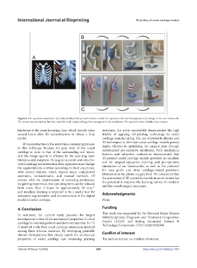
Figure 6. Pre-operative simulation. (A) Individualized 3D-printed silicone model. Pre-operative (B) and intraoperative (C) design of the ear framework.
The red arrows and dashed-line box mark the sixth costal cartilage that was spared in the simulation. The patient’s name is hidden by a mosaic.
limitation is the manufacturing time, which usually takes outcomes, the study successfully demonstrated the high
several hours after 3D reconstruction to obtain a final fidelity of applying 3D-printing technology to costal
model. cartilage manufacturing. The use of printable silicone and
3D reconstruction is the most time-consuming process 3D techniques to fabricate costal cartilage models proved
in this technique because the gray level of the costal highly effective in optimizing the surgical plan through
cartilage is close to that of the surrounding soft tissue, personalized pre-operative simulation. Both mechanical
and the image quality is affected by the scanning layer features and subjective evaluations demonstrated that
thickness and sharpness. To acquire accurate and objective 3D-printed costal cartilage models provided an excellent
costal cartilage reconstruction data, operators must change tool for surgical education, training, and pre-operative
the segmentation’s contrast according to their experience simulation of ear frameworks, as well as the potential
after several failures, which require many complicated for nose grafts and other cartilage-related prosthetic
automatic, semiautomatic, and manual methods. Of fabrication in the plastic surgery field. We considered that
course, with the improvement of operating proficiency the promotion of 3D-printable models in more centers has
by gaining experience, the operating time can be reduced the potential to improve the learning curves of residents
from more than 2 hours to approximately 30 min, and the overall surgery outcomes.
77
and machine learning is expected to be a useful tool for Acknowledgments
automatic segmentation and reconstruction of the digital
model of costal cartilage. None.
4. Conclusion Funding
In summary, the current study presents the largest This work was supported by the National Major Disease
investigation to date of the mechanical properties of costal Multidisciplinary Diagnosis and Treatment Cooperation
cartilage in microtia patients and demonstrates that the 75 Project [21025] and Beijing Municipal Science &
A material is the best costal cartilage simulation material Technology Commission [Z221100007422084].
among three silicone materials. By developing printable
silicone formulations that closely match the mechanical Conflict of interest
properties of costal cartilage and evaluating printing The authors declare no conflicts of interest.
Volume 10 Issue 1 (2024) 224 https://doi.org/10.36922/ijb.1007