
Page 102 - IJB-2-1
P. 102
Artificial vascularized scaffolds for 3D-tissue regeneration — a report of the ArtiVasc 3D Project
epidermal layer,
• To integrate the artificial vascular system pro-
duced in WP6 in the fatty tissue,
• To build up a vascularized composite tissue graft,
including a fat, dermal and epidermal layer,
• To characterize and evaluate cell and tissue
properties considering in particular morphology,
viability, cell proliferation and the expression of
specific markers.
6.3 Objectives for WP10
The overall aim of this work package was to validate
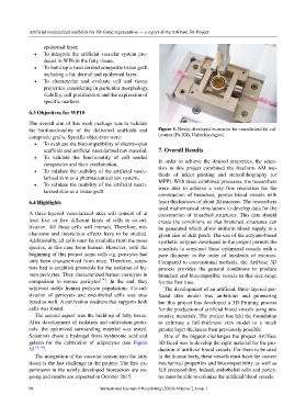
the biofunctionality of the delivered scaffolds and Figure 5. Newly developed bioreactor for vascularized fat cul-
composite grafts. Specific objectives were: tivation (Fh-IGB, Unitechnologies).
• To evaluate the biocompatibility of electro-spun
scaffolds and artificial vascularized raw material, 7. Overall Results
• To validate the functionality of cell seeded
composites and their combination, In order to achieve the desired properties, the scien-
• To validate the usability of the artificial vascu- tists in this project combined the freeform AM me-
larized skin as a pharmaceutical test system, thods of inkjet printing and stereolithography (or
• To validate the usability of the artificial vascu- MPP). With these combined processes, the researchers
larized skin as a tissue graft. were able to achieve a very fine resolution for the
construction of branched, porous blood vessels with
6.4 Highlights layer thicknesses of about 20 microns. The researchers
used mathematical simulations to develop data for the
A three-layered vascularized skin will consist of at construction of branched structures. This data should
least four or five different kinds of cells in co-cul- create the conditions so that branched structures can
tivation. All those cells will interact. Therefore, me- be generated which allow uniform blood supply to a
chanisms and interaction effects have to be studied. given size of skin patch. The use of the acrylate-based
Additionally, all cells must be available from the same synthetic polymer developed in the project permits the
species, in this case from human. However, until the scientists to construct these optimized vessels with a
beginning of this project some cells e.g. pericytes had pore diameter in the order of hundreds of microns.
only been characterized from mice. Therefore, scien- Compared to conventional methods, the ArtiVasc 3D
tists had to establish protocols for the isolation of hu- process provides the general conditions to produce
man pericytes. They characterized human pericytes in branched and biocompatible vessels in this size range
comparison to mouse pericytes [14] . In the end, they for the first time.
achieved stable human pericyte populations. Co-cul- The development of an artificial, three-layered per-
tivation of pericytes and endothelial cells was ana- fused skin model was ambitious and pioneering
lyzed as well. A cultivation medium that supports both but this project has developed a 3D Printing process
cells was found. for the production of artificial blood vessels using inn-
The second aspect was the build-up of fatty tissue. ovative materials. The project has laid the foundation
After development of isolation and cultivation proto- to cultivate a full-thickness skin model to a much
cols, the optimized surrounding material was tested. greater layer thickness than previously possible.
Scientists chose a hydrogel from hyaluronic acid and One of the biggest challenges the project ArtiVasc
gelatin for the cultivation of adipocytes (see Figure 3D faced was to develop the right material for the pro-
5) [15–19] . duction of artificial blood vessels. For them to be used
The integration of the vascular system into the fatty in the human body, these vessels must have the correct
tissue is the last challenge in the project. The first ex- mechanical properties and biocompatibility as well as
periments in the newly developed bioreactors are on- full processability. Indeed, endothelial cells and pericy-
going and results are expected in October 2015. tes must be able to colonize the artificial blood vessels.
98 International Journal of Bioprinting (2016)–Volume 2, Issue 1