
Page 358 - IJB-10-6
P. 358
International Journal of Bioprinting Collagen hydrolysate-loaded ODMA/PEGDMA scaffold
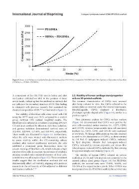
Figure 9. Stress–strain behaviors of polyethylene glycol dimethacrylate (PEGDMA) compared to PEGDMA with 10% oligomers of dopamine methacrylate
(ODMA) (i.e., ODMA–PEGDMA).
A comparison of the CH FTIR spectra before and after 3.5. Viability of human cartilage stem/progenitor
sterilization exhibited no shift in the position of these cells on 3D-printed scaffolds
amide bands, indicating that the sterilization methods did The common characteristics of CSPCs were accessed
not influence the secondary structure of CH. This finding after being cultured in vitro. The CSPCs adhered to the
is consistent with previous research that examined the culture plates as observed under the inverted microscope.
chemical composition of CH, 29,30 as illustrated in Figure 10. Morphologically, CSPCs displayed a fibroblastic
phenotype (spindle-shaped cells) (Figure 13), similar to a
The viability of fibroblast cells when tested with CH previous report. 34
using the MTT assay over 24 h compared to a control
group (without CH) yielded insightful results. The Flow cytometry analysis for CSPCs surface markers
fibroblast cells cultured in a medium containing 20% w/v (Figure 14) demonstrated that CSPCs were positive for
CH and post-sterilized by filtration, EtO, beta radiation, stem cells-associated surface markers (i.e., CD73, CD90,
and gamma radiation demonstrated survival rates of and CD105), whereas hematopoietic stem cell-associated
112.46%, 122.90%, 117.40%, and 118.95%, respectively. markers (i.e., CD34, CD45, and CD11b) were expressed
These results are illustrated in Figure 11. Furthermore, at low levels. Tri-lineage differentiation was also assessed
when the cells were stained with fluorescent markers to confirm the characteristics of CSPCs, as demonstrated
to assess viability within the CH-containing culture by Oil Red O (adipogenic-induced CSPCs; indicated
medium after various sterilization methods, the cells by lipid droplets), Alizarin Red (osteogenic-induced
exhibited a prominent green fluorescence from the CSPCs; indicated by calcium deposits), and Alcian Blue
calcein staining of fibroblast cells, which indicates a high (chondrogenic-induced CSPCs; indicated by blue staining
number of live cells. In contrast, the red stains of ethidium for glycosaminoglycans) staining (Figure 15).
staining revealed only a minimal presence of dead cells, The viability of human CSPCs cultured on various
consistent across all samples (Figure 12). These consistent scaffolds at 24, 48, and 72 h intervals was compared to the
results suggest that the sterilization methods did not control (culture plate) group (Figure 16). For PEGDMA
detrimentally affect cell viability, aligning with previous scaffolds, the survival rates were 72.89%, 56.60%, and
research on the cell toxicity of CH subjected to different 119.70% at the respective time points. When ODMA was
sterilization methods. 31–33 incorporated into the PEGDMA scaffold, the survival rates
Volume 10 Issue 6 (2024) 350 doi: 10.36922/ijb.4385