
Page 360 - IJB-10-6
P. 360
International Journal of Bioprinting Collagen hydrolysate-loaded ODMA/PEGDMA scaffold
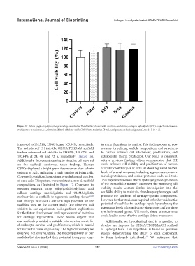
Figure 11. A bar graph displaying the percentage survival of fibroblasts cultured with medium containing collagen hydrolysate (CH) subjected to various
sterilization techniques, i.e., filtration (filter), ethylene oxide (EtO), beta radiation (beta), and gamma radiation (gamma), for 24 h (n = 3).
improved to 102.71%, 139.62%, and 163.34%, respectively. term cartilage tissue formation. This finding opens up new
The inclusion of CH into the ODMA/PEGDMA scaffold avenues for refining scaffold compositions and structures
further enhanced cell viability to 138.85%, 148.67%, and to further enhance cell attachment, proliferation, and
190.64% at 24, 48, and 72 h, respectively (Figure 16). extracellular matrix production. Our result is consistent
Additionally, fluorescent staining to visualize cell survival with a previous finding, which demonstrated that CH
on the scaffolds confirmed these findings. Human could enhance cell viability and proliferation of human
CSPCs displayed a bright green fluorescence after calcein articular chondrocytes in vitro via downregulated mRNA
staining at 72 h, indicating a high number of living cells. levels of several enzymes, including aggrecanases, matrix
Conversely, ethidium homodimer revealed a small number metalloproteinases, and serine proteases such as Htra1.
of dead cells. This pattern was consistent across all scaffold This may have beneficial effects by delaying the degradation
36
compositions, as illustrated in Figure 17. Compared to of the extracellular matrix. Moreover, the promising cell
previous research using polyglycolide/polylactic acid viability results warrant further investigation into the
cellular cartilage matrix/gelatin and ODMA/gelatin scaffolds’ ability to maintain chondrocyte phenotype and
methacrylate as scaffolds to regenerate cartilage tissue, 10,35 promote the synthesis of cartilage-specific components.
our findings indicated a similarly high potential for the However, further studies are required to further validate the
scaffolds used in the current study. The observed cell potential of scaffolds for cartilage repair by analyzing the
viability in our experiments had significant implications expression levels of chondrocyte phenotypes and cartilage
for the future development and improvement of materials synthesis-related genes. Ultimately, these advancements
for cartilage regeneration. These results suggest that could lead to more effective cartilage defect treatments.
our scaffolds provided a suitable microenvironment for Additionally, we hypothesized that it is possible to
chondrocyte survival and proliferation, which is crucial develop and improve the ODMA/PEGDMA/CH scaffold
for successful tissue engineering. The high cell viability we in hydrogel form. This hypothesis is based on previous
observed not only validated the biocompatibility of our studies demonstrating the ability of each component
scaffolds but also implied their potential to support long- to form hydrogels individually. We anticipate that
10
Volume 10 Issue 6 (2024) 352 doi: 10.36922/ijb.4385