
Page 32 - IJB-7-4
P. 32
3D-printed Bolus in Radiotherapy
conventional flat bolus with a uniform size can hardly skin and have good efficacy in radiotherapy, but have been
match the patient’s unique body geometry and allow limited in practical use due to the shortcomings, including
for repeatable setup for treatment [7,8] . Therefore, it is an the immaturity of printed materials, inconvenient
urgent desire to customize a bolus for fitting any skin preparation, and time-consuming preparation.
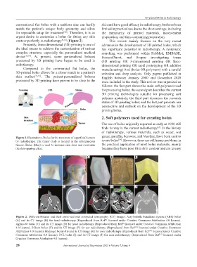
contour perfectly in radiotherapy [9-12] (Figure 2). This review mainly focuses on the very recent
Presently, three-dimensional (3D) printing is one of advances in the development of 3D printed bolus, which
the ideal means to achieve the customization of various has significant potential in radiotherapy. A systematic
complex structure, especially the personalized medical searching was performed within PubMed, EMBASE,
device [13,14] . At present, some personalized boluses ScienceDirect, and Scopus investigating terms
processed by 3D printing have begun to be used in (3D printing OR 3-dimensional printing OR three-
radiotherapy. dimensional printing OR rapid prototyping OR additive
Compared to the commercial flat bolus, the manufacturing) And (bolus OR polymers) with a careful
3D-printed bolus allows for a closer match to a patient’s selection and deep analysis. Only papers published in
skin surface [15,16] . The patient-personalized boluses English between January 2000 and December 2020
processed by 3D printing have proven to be close to the were included in the study. This review was organized as
follows: the first part shows the main soft polymers used
for processing bolus; the second part describes the current
3D printing technologies suitable for processing soft
polymer materials; the third part discusses the research
status of 3D printing bolus; and the last part presents our
perspective and outlook on the development of the 3D
printing bolus.
2. Soft polymers used for creating bolus
The use of bolus originally reported as early as 1920 still
finds its way in the current radiotherapy . In the history
[17]
of radiotherapy, various materials, such as water, wet
gauze, paraffin, beeswax, and Vaseline, have been used to
Figure 1. Illustration of bolus for the treatment of superficial tumors
[18]
by radiotherapy. The tumor (red) is located in the subcutaneous create bolus . However, there are still many problems in
tissues. Bolus (blue) is used to increase skin dose and overcome the practical application of most bolus materials, mainly
the skin-sparing effect. because they have poor fit to skin contour and are uneasy
A B C D E
F G H I J
Figure 2. Different boluses and their cross-sectional computed tomography (CT) images. Acrylonitrile butadiene styrene (ABS) bolus
(A) and its CT image (B) for head radiotherapy (Reproduced from Ref licensed under Creative Commons Attribution 4.0 license).
[8]
Agilus-60 bolus (C) and its CT image (D) for head radiotherapy (Reproduced from Ref licensed under Creative Commons Attribution
[9]
4.0 license). Silicon bolus (E) and its CT image (F) for ear radiotherapy (Reproduced from Ref licensed under Creative Commons
[10]
Attribution 4.0 license). Hydrogel bolus (G) and its CT image (H) for nose radiotherapy (Reproduced from Ref licensed under Creative
[10]
Commons Attribution 4.0 license). PCL bolus (I) and its CT image (J) for nose radiotherapy (Reproduced from Ref licensed under
[12]
Creative Commons Attribution 4.0 license).
28 International Journal of Bioprinting (2021)–Volume 7, Issue 4