
Page 139 - IJB-9-4
P. 139
International Journal of Bioprinting 3D-printed scaffolds for osteochondral defects
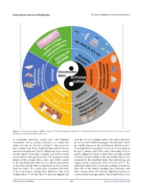
Figure 1. Schematic illustration of different aspects of 3D-printed gradient scaffolds for osteochondral defects in this review. (Some of the icons used in
this figure are derived from Biorender.com.)
of noncellular structures, usually only a few hundred cross the articular cartilage surface. The cells in this layer
nanometers, whose primary function is to reduce the are round and randomly arranged chondrocytes, which
surface friction on articular cartilage . This protective are usually known as the proliferating chondrocytes .
[10]
[11]
layer contains high levels of glycoproteins, also known as From superficial to deep layers, there is an increasing trend
mucosal proteoglycans. Type II collagen and water content in type I collagen and GAGs, and a decreasing trend in
are the highest, while type I collagen and GAGs content type II collagen and water content. Due to its high content
are the lowest in the superficial layer. The arrangement and of GAGs, the permeability of the intermediate layer is low
density of the collagen fibers, water, and GAGs content compared to the superficial layer, thus moderating and
in the superficial layer allow for the highest permeability supporting the compressive stress from the joint . The
[12]
of this layer and the effective dispersion of shear stresses radial layer (deep layer) of articular cartilage accounts for
from the joints. The middle layer comprises 40%–60% 20%–50% of the hyaline cartilage layer thickness, with
of the total hyaline cartilage layer thickness, with thick thick collagen fibers (60–140 nm) aligned perpendicular
collagen fibers (9–60 nm) that are unevenly aligned and to the articular cartilage surface. The chondrocytes in this
Volume 9 Issue 4 (2023) 131 https://doi.org/10.18063/ijb.724