
Page 140 - IJB-9-4
P. 140
International Journal of Bioprinting 3D-printed scaffolds for osteochondral defects
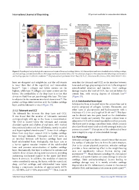
Figure 2. Physiological and pathological occurrence and repair of bone and cartilage defects. (A) Diseased joint and osteochondral units including cartilage,
calcified cartilage, and subchondral bone. (B) Categoriesofosteochondral defect. (C) The schematic diagram of the osteochondral physiologic environment
and healing capacities in different conditions (Reproduced with permission from Huey DJ, Hu JC, Athanasiou KA, Science, 2012, 338(6109):917–921).
[92]
layer are elongated and subglobular, and the cell density seen that the tidemark and CCZ, as the interface between
is lower than that of the superficial and intermediate bone and cartilage, play an important role in the integrated
layers . Type I collagen and GAGs content are the osteochondral structure and function. Total cartilage
[13]
highest, while type II collagen and water content are the damage reaches the level of CCZ, but not yet below the
lowest. The permeability of the deep layer is so low that cement line, with varying degrees of tidemark loss
[18]
almost no fluid flow can pass through this layer. This layer (Figure 2B).
is subjected to the maximum interfacial shear stress . The
[14]
partial cartilage defect involves only the hyaline cartilage 2.1.3. Subchondral bone layer
layer, and the tidemark is intact (Figure 2B). Subchondral bone is located below the cement line and is
mainly composed of collagen, laminin, fibronectin, and
2.1.2. Tidemark and CCZ other types of glycoproteins and hydroxyapatite with a
[18]
The tidemark lies between the deep layer and CCZ. thickness of 2–5 nm and length of 20–80 nm . This layer
It was found that the number of tidemarks increased can be divided into two parts based on the distribution
correspondingly with age as the tissue is reconstructed. of blood vessels and porosity. The upper cortical bone is
The CCZ is located below the tidemark and contains adjacent to CCZ with minimal vascularity and low porosity.
abundant apatite and alkaline phosphatase, with marked The lower part is spongy cancellous bone, which contains
tissue mineralization and low cell density, mostly rounded abundant blood vessels and randomly arranged trabecular
[19]
and hypertrophied chondrocytes . Some thick collagen porous structures . Disruption of the subchondral bone
[15]
fibers from deep layer connect CCZ to hyaline cartilage layer integrity is a sign of osteochondral damage.
layer through tidemark. Tidemarks and CCZ serve as 2.2. Pathology
an interface between soft hyaline cartilage layer and Articular cartilage is an elastic, smooth tissue that encases
hard subchondral bone. Biologically, this layer acts as the surface of articular bone to form the joint structure.
a barrier against vascular invasion of the subchondral Due to its unique physical properties, articular cartilage
bone and prevents mineralization of hyaline cartilage provides a force cushioning effect in the weight-bearing
layer. Mechanically, this layer is subjected to extreme and area and significantly reduces friction during joint
variable shear stresses during joint movement, providing movements [20,21] . Many diseases including OA, rheumatoid
cushioning mechanical support to the upper and lower arthritis, and sports injuries can cause damage to articular
layers it connects. In addition, the modulus of elasticity cartilage. Their pathophysiological factors leading to
varies considerably among the layers, with the superficial, cartilage damage vary: it is mainly articular cartilage
deep, calcified cartilage, and subchondral bone layers degeneration in OA, inflammatory erosion in rheumatoid
having a compressive modulus of approximately 0.079, arthritis (RA), and mechanical abrasion in sports injury .
[22]
2.1, 320 MPa, and 5.7 GPa, respectively [16,17] . Thus, it can be
Volume 9 Issue 4 (2023) 132 https://doi.org/10.18063/ijb.724