
Page 145 - IJB-9-4
P. 145
International Journal of Bioprinting 3D-printed scaffolds for osteochondral defects
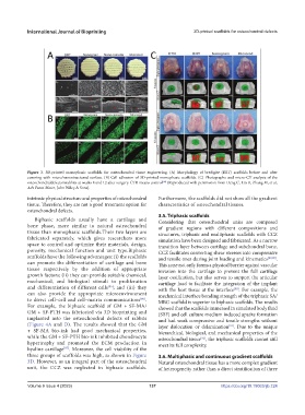
Figure 3. 3D-printed monophasic scaffolds for osteochondral tissue engineering. (A) Morphology of bredigite (BRT) scaffolds before and after
covering with micro/nanostructured surface. (B) Cell adhesion of 3D-printed monophasic scaffolds. (C) Photographs and micro-CT analysis of the
osteochondraldefectsinrabbits at weeks 8 and 12 after surgery. CTR means control (Reproduced with permission from Deng C, Lin R, Zhang M, et al,
[84]
Adv Funct Mater, John Wiley & Sons).
intrinsic physical structure and properties of osteochondral Furthermore, the scaffolds did not show all the gradient
tissue. Therefore, they are not a good treatment option for characteristics of osteochondral tissues.
osteochondral defects.
3.5. Triphasic scaffolds
Biphasic scaffolds usually have a cartilage and Considering that osteochondral units are composed
bone phase, more similar to natural osteochondral of gradient regions with different compositions and
tissue than monophasic scaffolds.Their two layers are structures, triphasic and multiphasic scaffolds with CCZ
fabricated separately, which gives researchers more simulation have been designed and fabricated. As a narrow
space to control and optimize their materials, design, transition layer between cartilage and subchondral bone,
porosity, mechanical function and unit type.Biphasic CCZ facilitates converting shear stresses into compressive
scaffolds have the following advantages: (i) the scaffolds and tensile ones during joint loading and kinematics [88,89] .
can promote the differentiation of cartilage and bone This zone not only forms a physical barrier against vascular
tissue respectively by the addition of appropriate invasion into the cartilage to prevent the full cartilage
growth factors; (ii) they can provide suitable chemical, layer ossification, but also serves to support the articular
mechanical, and biological stimuli to proliferation cartilage load to facilitate the integration of the implant
and differentiation of different cells [85] ; and (iii) they with the host tissue at the interface . For example, the
[90]
can also provide the appropriate microenvironment mechanical interface bonding strength of the triphasic SA/
to direct cell–cell and cell–matrix communications [86] . MBG scaffold is superior to biphasic scaffolds. The results
For example, the biphasic scaffold of GM + SF-MA/ showed that the scaffolds immersed in simulated body fluid
GM + SF-PTH was fabricated via 3D bioprinting and (SBF) and cell culture medium induced apatite formation
implanted into the osteochondral defects of rabbits and had weak compressive and tensile strengths without
(Figure 4A and D). The results showed that the GM layer dislocation or delamination . Due to the unique
[91]
+ SF-MA bio-ink had good mechanical properties, hierarchical, biological, and mechanical properties of the
while the GM + SF-PTH bio-ink inhibited chondrocyte osteochondral tissue , the triphasic scaffolds cannot still
[92]
hypertrophy and promoted the ECM production in meet its full complexity.
hyaline cartilage [87] . Moreover, the cell viability of the
three groups of scaffolds was high, as shown in Figure 3.6. Multiphasic and continuous gradient scaffolds
3D. However, as an integral part of the osteochondral Natural osteochondral tissue has a more complex gradient
unit, the CCZ was neglected in biphasic scaffolds. of heterogeneity rather than a direct stratification of three
Volume 9 Issue 4 (2023) 137 https://doi.org/10.18063/ijb.724