
Page 253 - IJB-9-4
P. 253
International Journal of Bioprinting Applications of 3D printing in aging
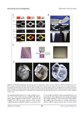
Figure 4. (A) Multiangle photographs of 3D-printed structures of three different types of vascular constructs . Reproduced with permission from
[18]
IOP Publishing, Copyright © 2022, IOP Publishing. (B) 3D-printed model of the patient (top). Calcification of the aortic valve leaflet (red arrow) [143] .
Reproduced with permission from Wolters Kluwer Health, Copyright © 2015, Wolters Kluwer Health. (C) 3D printing of a personalized heart patch.
From left to right: a 3D model of the heart patch, printing method, and heart patch with printed blood vessels [145] . Reproduced under Creative Commons
license. (D) 3D-printed collagen heart [149] . Reproduced with permission from The American Association for the Advancement of Science, Copyright
© 2022, AAAS.
the urgently needed structures for cardiovascular diseases. tensile strength and elastic modulus comparable to those of
Wu et al. [146] 3D-printed vascular scaffolds with DIW by native arteries. The scaffold aided in the supply of nutrients
using gelatin–alginate–montmorillonite nanocomposite and cellular infiltration. The hemolysis rate of the scaffold
bioinks. The inner and outer surfaces of the vascular also fulfilled the benchmark for vascular replacement,
scaffold displayed networked microporous structures with and the scaffold’s rupture pressure was similar to that of
Volume 9 Issue 4 (2023) 245 https://doi.org/10.18063/ijb.732