
Page 249 - IJB-9-4
P. 249
International Journal of Bioprinting Applications of 3D printing in aging
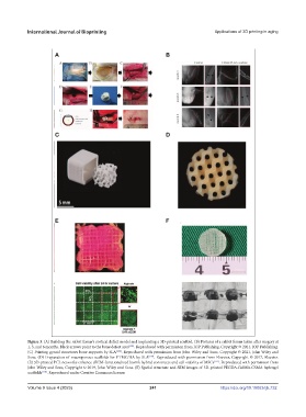
Figure 3. (A) Building the rabbit femur’s cortical defect model and implanting a 3D-printed scaffold. (B) Pictures of a rabbit femur taken after surgery at
1, 3, and 6 months. Black arrows point to the bone defect area [105] . Reproduced with permission from IOP Publishing, Copyright © 2021, IOP Publishing.
(C) Printing gyroid structures bone supports by SLA [106] . Reproduced with permission from John Wiley and Sons, Copyright © 2021, John Wiley and
Sons. (D) Preparation of macroporous scaffolds for PTMC/HA by SLA [107] . Reproduced with permission from Elsevier, Copyright © 2017, Elsevier.
(E) 3D-printed PCL networks enhance cECM-functionalized bioink hybrid constructs and cell viability of MSCs [117] . Reproduced with permission from
John Wiley and Sons, Copyright © 2019, John Wiley and Sons. (F) Spatial structure and SEM images of 3D-printed PEGDA-GelMA-CSMA hydrogel
scaffolds [118] . Reproduced under Creative Commons license.
Volume 9 Issue 4 (2023) 241 https://doi.org/10.18063/ijb.732