
Page 225 - IJB-9-5
P. 225
International Journal of Bioprinting Hydrogels for 3D bioprinting
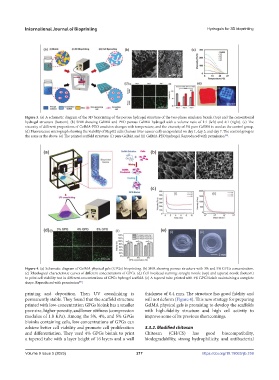
Figure 3. (a) A schematic diagram of the 3D bioprinting of the porous hydrogel structure of the two-phase emulsion bioink (top) and the conventional
hydrogel structure (bottom). (b) SEM showing GelMA and PEO porous GelMA hydrogel with a volume ratio of 1:1 (left) and 4:1 (right). (c) The
viscosity of different proportions of GelMA-PEO emulsion changes with temperature, and the viscosity of 5% pure GelMA is used as the control group.
(d) Fluorescence micrograph showing the viability of HepG2 cells (human liver cancer cell) encapsulated on day 1, day 3, and day 7. The control group is
[35]
the same as the above. (e) The printed scaffold structure: (i) pure GelMA and (ii) GelMA-PEO hydrogel. Reproduced with permission .
Figure 4. (a) Schematic diagram of GelMA physical gels (GPGs) bioprinting. (b) SEM showing porous structure with 3% and 5% GPGs concentration.
(c) Rheological characteristic curves of different concentrations of GPGs. (d) Cell live/dead staining: straight nozzle (top) and tapered nozzle (bottom)
to print cell viability test in different concentrations of GPGs hydrogel scaffold. (e) A tapered tube printed with 4% GPG bioink maintaining a complete
shape. Reproduced with permission .
[85]
printing and deposition. Then UV crosslinking is thickness of 0.4 mm. The structure has good fidelity and
permanently stable. They found that the scaffold structure will not deform (Figure 4). This new strategy for preparing
printed with low-concentration GPGs bioink has a smaller GelMA physical gels is promising to develop the scaffolds
pore size, higher porosity, and lower stiffness (compression with high-fidelity structure and high cell activity to
modulus of 1.8 KPa). Among the 3%, 4%, and 5% GPGs improve some of its previous shortcomings.
bioinks containing cells, low concentrations of GPGs can
achieve better cell viability and promote cell proliferation 3.3.2. Modified chitosan
and differentiation. They used 4% GPGs bioink to print Chitosan (CH/CS) has good biocompatibility,
a tapered tube with a layer height of 16 layers and a wall biodegradability, strong hydrophilicity, and antibacterial
Volume 9 Issue 5 (2023) 217 https://doi.org/10.18063/ijb.759