
Page 227 - IJB-9-5
P. 227
International Journal of Bioprinting Hydrogels for 3D bioprinting
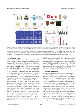
Figure 5. (A) (i) Bioprinting a scaffold with PACG-GelMA hydrogel-Mn as the top layer and PACG-GelMA hydrogel-BG as the bottom layers;
2 +
(ii) schematic diagram of repairing osteochondral defects. Reproduced with permission [100] . (B) 3D bioprinting of PEG-SA-nano clay hydrogel. (i) 3D
bioprinting of different shapes: hollow cubes, hemispheres, pyramids, twisted ear shapes, and noses; (ii) meshes with tough and biocompatible hydrogels;
(iii) live and dead staining of HEK cells; (iv) HEK cell viability at 7 day. Reproduced with permission . (C) (i) Infrared spectrum: hydrogel (black line)
[53]
and hydrogel attached to GO-np (red line); (ii) chondrocytes in hydrogel, GO-np, hydrogel/GO–vitality test of np, and blank group. Reproduced with
permission [105] . (iii) Swelling ratio of printing and (iv) porosity of Gel-SA hydrogel scaffold containing CNTs. Reproduced with permission [106] .
4.1.1. Bioactive glass and differentiation of hBMSCs. After being implanted in
Bioactive glass (BG) is a silicate glass composed of SiO , the body for a period of time, it shows excellent repair
2
Na O, CaO, etc. It has good biocompatibility, bioactivity, and performance for osteochondral defects. Also, regenerated
2
degradability. The elements released during degradation cartilage and subchondral bone were apparently observed
can form a hydroxyapatite (HA) layer on the surface of in the rat model. Therefore, in the process of bone repair
the material similar to natural bone, which then forms and regeneration, BG showed obvious proliferation and
a strong chemical bond with the adjacent bone surface differentiation effects on hBMSCs cells (Figure 5A).
and can promote the formation of new bone . Ding
[98]
et al. fabricated a framework-filled structure consisting 4.1.2. Hydroxyapatite (HA)
[99]
of polylactic acid (PLA), BG, and bone cement. The results HA is the main inorganic component of animal bones, so it
showed that the scaffold provides instant and stable fixation is widely applied in hard tissue engineering such as bones
of the bone defect. It is also characterized by continuous and teeth. Aihemaiti et al. [101] optimized the construction
osteogenesis induction. The sustained degradation of parameters of 3D PLA/HA composite bone plates
PLA + 1% BG composite scaffold (PBG) provides space through many experimental tests and analyzed the effect
for bone growth. Gao et al. [100] used the 3D bioprinting of of HA content on the flexural strength of the specimens.
poly (N-acryloyl 2-glycine) (PACG) and GelMA hydrogel When the HA content is 20%, the cross-section is rough
to obtain a biodegradable and supramolecular hydrogen compared to pure PLA (0% HA) and 10% HA specimens.
bond-enhanced crosslinked gelatin hydrogel scaffold. To In addition, it contains a large amount of dents and pores,
better promote osteochondral regeneration, they added which reduces the bending properties of the specimens.
biologically active manganese ions (Mn ) and BG to Ergul et al. bioprinted CH/PVA scaffolds containing HA
2+
the scaffold materials, forming PACG-GelMA hydrogel- and tested the performance of HA scaffolds with different
Mn as the top layer while PACG-GelMA hydrogel-BG ratios [102] . They found that 15 wt% HA and CH/PVA
2+
is the bottom support. The scaffold was applied to living hydrogels have obvious advantages as bioinks. The elastic
cartilage repair, and it was found that the incorporation modulus of the scaffold can reach 91.14 MPa, which is
of BG improved the proliferation and differentiation of close to the elastic modulus of natural bone. Then, bone
hBMSCs. Loaded Mn can promote the chondrogenesis morphogenetic protein-2 (BMP-2) was inoculated onto
2+
Volume 9 Issue 5 (2023) 219 https://doi.org/10.18063/ijb.759