
Page 11 - IJB-9-6
P. 11
International Journal of Bioprinting Electrospinning PETG
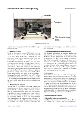
Figure 1. Electrospinning setup
systems on the Teas graph, and then the solubility region diameter. For each mesh type, a total of 9 measurements
was constructed. were considered.
2.3. Mesh fabrication 2.5. Chemical composition characterization
Based on the solubility results, PETG (20% w/v) was The chemical composition of produced meshes was
dissolved in different splits of DCM/TFA (85/15, analyzed using Fourier-transform infrared spectroscopy
70/30, 60/40, 50/50, 40/60, 30/70, 15/85 [% v/v]), and (FTIR) and Raman spectroscopy. FTIR was conducted
electrospun using solution electrospinning (Spraybase, using Varian 670-IR spectrometer (Agilent Technologies,
Ireland) (Figure 1). Meshes were produced at room CA, USA). Each sample was scanned 20 times at the
temperature with a voltage of 16 kV, a feed flow rate of resolution of 1 cm , over a frequency scanning range
-1
4 mL/h, a humidity of 45% (±5%), and a distance between from 800 to 3100 cm . Raman spectroscopy was carried
-1
the needle tip and the collector, which was covered with out using Renishaw inVia confocal Raman microscope
aluminum foil, of 150 mm. Finally, the obtained meshes (Renishaw Plc., Gloucestershire, UK) using laser (532 nm)
were dried in vacuum for 48 hours to ensure the complete with a grating of 1200 g/mm in a regular mode and use of
removal of solvents. These were considered the optimized 50× magnification on the microscope.
parameters. Voltage values lower than 16 kV did not
enable the formation of a stable jet but only caused a 2.6. Inoculation
dropping effect, while values higher than 16 KV induced The ability of the electrospun meshes to sustain pathogen
electrospraying. Moreover, flow rate values lower than attachment and germination was assessed using the yellow
4 mL/h induced a fast evaporation of the solvent blocking rust spores’ inoculation process. The meshes were placed in
the needle, while values above 4 mL/h caused a dropping a tray in a stainless-steel pipe, simulating a wind tunnel. The
effect. spores were released in the pipe, landing on the meshes at
the bottom of the pipe. The meshes were kept in the pipe
2.4. Morphological analysis for 15–30 min, ensuring that all spores have settled on the
The morphology of produced meshes was characterized surfaces. Then, the inoculated surfaces were imaged under a
using the scanning electron microscopy (SEM). For SEM, light microscope to make sure that the spores were attached.
Quanta 650 (FEI company, Hillsboro, Oregon, USA) was Finally, the meshes were placed in the Innova 44 incubator
used, all meshes were sputter coated with gold/palladium (Eppendorf, Hamburg, Germany) set at 7°C for 24 h.
(Au/Pd (80/20)) using Quorum sputter coater (Quorum
tech, east Sussex, UK). Imaging was carried out using 3. Results and discussion
an acceleration voltage of 15 kV. The obtained images
were analyzed using the ImageJ software (Laboratory for 3.1. Solubility and electrospinnability
Optical and Computational Instrumentation, University Solubility, which describes how easy it is to dissolve
of Wisconsin, WI, USA), allowing to determine fiber the polymer in a solvent, and electrospinnability,
Volume 9 Issue 6 (2023) 3 https://doi.org/10.36922/ijb.0024