
Page 338 - v11i4
P. 338
International Journal of Bioprinting GradGelMA 3D-bioprinted vascular skin
repeatedly dropping a low-concentration gelatin solution optimizing the culture parameters for epidermal cells,
containing keratinocytes, ultimately resulting in a pre- dermal cells, and vascular endothelial cells, we prepared
vascularized multi-layered skin substitute. Despite these a hydrogel-based, cell-compatible composite ink for VS.
advances, significant challenges remain. Many studies Using 3D extrusion printing technology, we constructed
primarily assess endothelial cell viability without fully a stable in vitro dermal VS substitute. Additionally, the
characterizing in vitro microvascular network formation 3D-printed VS was implanted into skin defect models to
or functionality. Effective vascularization is critical for further investigate its reparative effects on damaged tissue.
nutrient delivery, oxygenation, and tissue integration, yet We believe this study provides a promising skin substitute
current methods often fail to recapitulate the hierarchical for clinical applications and offers valuable insights for
vascular networks found in native skin. future research.
Therefore, this study aimed to construct a multi-
layered skin substitute with a vascularized structure. First, 2. Methods
we modified gelatin with methacrylic anhydride to obtain 2.1. Three-dimensional bioprinting equipment
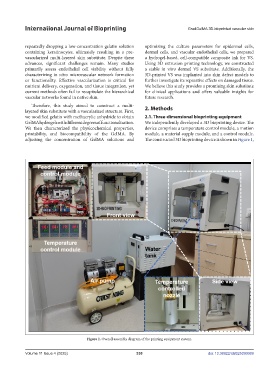
GelMA hydrogels with different degrees of functionalization. We independently developed a 3D bioprinting device. The
We then characterized the physicochemical properties, device comprises a temperature control module, a motion
printability, and biocompatibility of the GelMA. By module, a material supply module, and a control module.
adjusting the concentration of GelMA solutions and The constructed 3D bioprinting device is shown in Figure 1,
Figure 1. Overall assembly diagram of the printing equipment system.
Volume 11 Issue 4 (2025) 330 doi: 10.36922/IJB025090069