
Page 112 - OR-1-1
P. 112
A
D
B
C
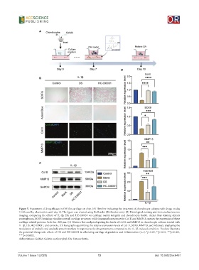
Figure 5. Assessment of drug efficacy in OA-like cartilage-on-chip. (A) Timeline indicating the treatment of chondrocyte cultures with drugs on day
7, followed by observation until day 10. The figure was created using BioRender (BioRender.com). (B) Histological staining and immunofluorescence
imaging, comparing the effects of IL-1β, DS, and HC-030031 on cartilage matrix integrity and chondrocyte health. Alcian blue staining detects
proteoglycans, SO/FG staining visualizes overall cartilage structure, while immunofluorescence for Col II and MMP13 assesses the expression of these
cartilage-related proteins. Scale bar: 200 μm. (C) Western blot analysis depicting the levels of Col II and MMP13 in chondrocyte cultures treated with
IL-1β, DS, HC-030031, and controls. (D) Bar graphs quantifying the relative expression levels of Col II, SOX9, MMP13, and Adamts5, displaying the
modulation of anabolic and catabolic protein markers in response to the drug treatments compared to the IL-1β-induced condition. The data illustrates
the potential therapeutic effects of DS and HC-030031 in alleviating cartilage degradation and inflammation (n=3, *p<0.05, **p<0.01, ***p<0.001,
****p<0.0001).
Abbreviations: GelMA: Gelatin methacryloyl; OA: Osteoarthritis.
Volume 1 Issue 1 (2025) 13 doi: 10.36922/or.8461