
Page 108 - OR-1-1
P. 108
A
C
B
D
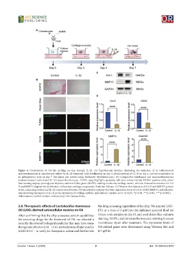
Figure 4. Construction of OA-like cartilage-on-chip through IL-1β. (A) Experimental timeline illustrating the induction of an inflammatory
microenvironment in chondrocyte culture by IL-1β treatment, with stabilization on day 3, administration of IL-1β on day 4, and test progression to
an inflammatory state on day 7. The figure was created using BioRender (BioRender.com). (B) Comparative histological and immunofluorescence
analyses between control and IL-1β-treated chondrocytes. TUNEL assay highlights apoptotic cells (red arrows indicate TUNEL-positive cells), alcian
blue staining displays proteoglycan deposits, safranin O/fast green (SO/FG) staining reveals the cartilage matrix, whereas immunofluorescence for Col
II and MMP13 displays the distribution of these key cartilage components. Scale bar: 200 μm. (C) Western blot detection of Col II and MMP13 protein
levels, comparing control and IL-1β-treated chondrocytes. (D) Quantitative analysis of protein expression levels of Col II, SOX9, MMP13, and Adamts5,
demonstrating the impact of IL-1β on the expression of cartilage anabolic and catabolic markers (n=3, *p<0.05, **p<0.01, ***p<0.001, ****p<0.0001).
Abbreviations: GelMA: Gelatin methacryloyl; OA: Osteoarthritis.
2.8. Therapeutic effects of Lactobacillus rhamnosus the drug screening capabilities of the chip. We injected LGG-
GG (LGG)-derived extracellular vesicles on OA EVs at a dose of 5 μM into the inflamed synovial fluid for
After confirming that the chip possesses certain capabilities 3 days, took samples on day 10, and used alcian blue-safranin
for screening drugs for the treatment of OA, we selected a staining, SO/FG, and immunofluorescence staining to assess
recently discovered biological medicine that may have some microtissue repair after treatment. The expression levels of
therapeutic effects on OA – LGG-derived extracellular vesicles OA-related genes were determined using Western blot and
(LGG-EVs) – to verify its therapeutic action and further test RT-qPCR.
Volume 1 Issue 1 (2025) 9 doi: 10.36922/or.8461