
Page 104 - OR-1-1
P. 104
A
B
C
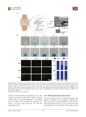
Figure 2. Design scheme and characterization of cartilage-on-chip. (A) Schematic representation of the cartilage-on-chip, displaying the placement of
chondrocytes within a GelMA hydrogel scaffold inside the culture medium and the corresponding experimental setup. The figure was created using
BioRender (BioRender.com). (B) The 96-h dye penetration image displays the interaction and stabilization process of the GelMA hydrogel with the
medium. (C) Fluorescence microscopy images on days 3, 6, and 10 of culture displaying the live/dead chondrocytes within the scaffolds (C, left).
Quantitative bar graph depicts the percentage of live/dead cells over time (C, right), indicating cell viability and proliferation within the GelMA matrix
(n=3). Scale bar: 400 μm.
Abbreviation: GelMA: Gelatin methacryloyl.
washed 3 times with TBST, each wash lasting 10 min. The 2.5.3. Real-time polymerase chain reaction
membrane was then incubated with a diluted secondary The excess culture medium was removed using PBS, and
antibody solution while gently shaking for 1.5 h, followed 200 μL of total RNA extraction reagent was added to the
by three 10-min washes with TBST. The membrane was cells. The mixture was then incubated at 4°C for 15 min.
imaged accordingly using ChemiDoc MP (Bio-Rad The lysate was collected into a 1.5 mL centrifuge tube, and
Laboratories, USA). 40 μL of chloroform was added. The mixture was vortexed
Volume 1 Issue 1 (2025) 5 doi: 10.36922/or.8461