
Page 70 - OR-1-1
P. 70
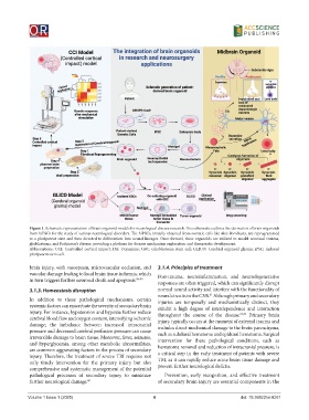
Figure 1. Schematic representation of brain organoid models for neurological disease research. This schematic outlines the derivation of brain organoids
            from hiPSCs for the study of various neurological disorders. The hiPSCs, initially obtained from somatic cells like skin fibroblasts, are reprogrammed
            to a pluripotent state and then directed to differentiate into neural lineages. Once formed, these organoids are utilized to model neuronal trauma,
            glioblastoma, and Parkinson’s disease, providing a platform for disease mechanism exploration and therapeutic development.
            Abbreviations: CCI: Controlled cortical impact; DA: Dopamine; GSC: Glioblastoma stem cell; GLICO: Cerebral organoid glioma; iPSC: Induced
            pluripotent stem cell.


            brain injury, with vasospasm, microvascular occlusion, and   3.1.4. Principles of treatment
            vascular damage leading to local brain tissue ischemia, which   Post-trauma,  neuroinflammation,  and  neurodegenerative
            in turn triggers further neuronal death and apoptosis. 56-59  responses are often triggered, which can significantly disrupt

            3.1.3. Homeostasis disruption                     normal neural activity and interfere with the functionality of
                                                              neural circuits in the CNS.  Although primary and secondary
                                                                                  61
            In addition to these pathological mechanisms, certain   injuries are temporally and mechanistically distinct, they
            systemic factors can exacerbate the severity of secondary brain   exhibit a high degree of interdependence and interaction
            injury. For instance, hypotension and hypoxia further reduce   throughout the course of the disease. 62,63  Primary brain
            cerebral blood flow and oxygen content, intensifying ischemic   injury typically occurs at the moment of external trauma and
            damage; the imbalance between increased intracranial   includes direct mechanical damage to the brain parenchyma,
            pressure and decreased cerebral perfusion pressure can cause   such as subdural hematoma and epidural hematoma. Surgical
            irreversible damage to brain tissue. Moreover, fever, seizures,   intervention for these pathological conditions, such as
            and hyperglycemia, among other metabolic abnormalities,   hematoma removal and reduction of intracranial pressure, is
            are common aggravating factors in the process of secondary
            injury. Therefore, the treatment of severe TBI requires not   a critical step in the early treatment of patients with severe
            only timely intervention for the primary injury but also   TBI, as it can rapidly reduce acute brain tissue damage and
            comprehensive and systematic management of the potential   prevent further neurological deficits.
            pathological  processes of secondary  injury to  minimize   Prevention, early recognition, and effective treatment
            further neurological damage. 60                   of secondary brain injury are essential components in the


            Volume 1 Issue 1 (2025)                         4                                 doi: 10.36922/or.8261